ISSN: 0970-938X (Print) | 0976-1683 (Electronic)
Biomedical Research
An International Journal of Medical Sciences
Research Article - Biomedical Research (2016) Volume 27, Issue 4
Preprocessing digital breast mammograms using adaptive weighted frost filter
1Deanship of Scientific Research, King Saud University Riyadh Saudi Arabia
2Faculty of Computing, Universiti Teknologi Malaysia
3Department of Computer Science, Imam Muhammad Bin Saud Islamic University, Saudi Arabia
- *Corresponding Author:
- Dr. Muhammad Talha
Deanship of Scientific Research
King Saud University
Saudi Arabia
Accepted date: April 27, 2016
Mammography became the most efficient tool for early detection among breast cancer patients because it can detect cancer up to two years earlier than a mass can be shown. Pre-processing and postprocessing of mammographic images involves high computational cost. Preprocessing is an essential element of any imaging modalities whose foremost aim is to execute such processes which can bring the image to that quality where it is suitable for further analysis and extraction of significant data. This paper talks about pre-processing which has great significance in mammographic image analysis due to poor quality of mammograms since they are captured at low dose of radiation while the high amount of radiation may jeopardize the patient’s health. Many techniques have been used to enhance image quality, image smoothing and noise restoration. The experimental results conclude that the proposed Adaptive Weighted Frost filter is the best suitable choice for eliminating noise from mammographic images and performs better comparatively. The comparison of proposed technique with various existing techniques is performed both qualitatively and quantitatively. The experiments have demonstrated that the proposed technique provides better results as compare to existing techniques.
Keywords
Mammograms, Enhancement, Frost filter, PSNR.
Introduction
Breast cancer is the uncontrolled growth of breast cells. Various aspects are connected with breast cancer such as genetics, age variation, obesity, asymmetry in breasts, ethnicity, history of breast feeding and menses [1]. A mammogram is a breast x-ray that is suggestive by the physician when there is something suspected [2]. The method contains the compression of breast between two plates with low dose of radiation because higher dose of radiation can destroy the patient’s health [3]. Screening mammography is performed for detecting breast cancer before it occurs and reduces the death rate. Only the early diagnosis is the approach to avoid mastectomy, reduce the likelihood to return, and increase the survival rates. Diagnostic mammography is conducted when a woman has signs of malignancy such as a mass which can be felt. Recent guidelines from the American Cancer Society, the AMA (American Medical Association), Health and Human Services Department of U.S and the ACR (American College of Radiology) advise screening mammography yearly for women, beginning at age 40. Various studies have verified that mammography performed annually reduced the rate of mortalities and are highly helpful in the early detection of breast cancers among women between 40 to 74 ages where the probability of treatment is very high [4-6].
In developing countries, breast cancer is frequently identified cancer and major source of fatality amongst women. Mammography provides the complete information regarding shape, structure and pathologies of breast which helps out in diagnosing breast cancer [7]. The complexity occurs while detecting masses from mammograms due to the similarities between the masses of normal and abnormal (benign or malignant) breast tissues [8]. The mammograms which are confirmed as abnormal after the results of mammography diagnostic are clinically divided into two classes: benign (noncancerous) and malignant (cancerous) [9,10]. It is also significant at this time to depict about the distinctiveness and features of mammograms which clinically distinguish between benign and malignant. The most important characteristics which tell either a mass is benign or malignant are its shape (round, irregular, etc) and margins (circumscribed, ill-defined, speculated, etc) [11,12]. The tumors with regular shape like round and oval frequently indicate a benign change whereas irregular shaped masses normally tells about malignancy as it is illustrated in Figure 1 [13].

Figure 1: Morphologic spectrum of masses [13].
Sometimes it becomes quiet complicated to distinguish between benign and malignant masses because of low-contrast. So it is pertinent to apply any automatic technique to enhance the visual quality of mammographic image in order to forefront the malignancies, if exists. Enhancing the contrast, extracting the features and removing the noise are the fundamental issues in mammograms which need to be addressed [14]. Noise may be expressed as any change in the mammogram that do not relate to variations in the X-ray attenuation of the object being imaged. X-ray quantum noise is the significant type of noise which is denoted by a Poisson distribution [15]. Such type of noise is generally introduced during mammograms image acquisition because of less number of x-ray photons. It reduces the contrast of the mammogram image mainly which results in the low value of signal-to-noise ratio (SNR) [16]. Resultantly, the contrast of mammograms becomes low because of quantum noise whose detection is not only a complicated job but also challenging for the radiologists. False-negatives for radiologist mammography interpretation vary from 10 to 30% due to different factors [17].
Some existing methods on image enhancement are median filtering, Frost filter, Wiener filter, histogram equalization and contrast limited adaptive histogram equalization. The radiologist uses various softwares like “ImageChecker” and “SecondLook” to enhance and/or zoom the selected part of the mammograms which are usually based on various enhancement, segmentation/cropping and zooming techniques. For the enhancement of mammograms, Bhateja et al. [18] used median filter to suppress salt and pepper noise. For noise restoration, the average of three filters (Frost, Adaptive Wiener and non-local mean) was proposed by Naveed et al. [19]. The mean value of these filters was replaced with noisy pixels. The authors implemented their technique on salt & pepper and quantum noise. They used PSNR and SSIM (structural similarity index measure) as performance measures to evaluate the performance of the proposed technique with various existing methods in literature. Numerous computer aided techniques for image processing have been developed and applied by many of researchers for the enhancement of mammographic images using various filters. In our proposed method, the Frost filter is modified which intelligently remove the noise from the mammograms. The details are discussed in the materials and methods section.
Materials and Methods
Adaptive weighted frost filter for quantum noise removal
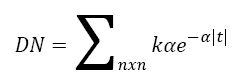
The quantum noise is cause due to uneven strike of photons on screen. During the attainment of images, this noise is usually introduced because of low count X-Ray photons. Since the existence of noise is directly proportional to degree of unevenness in quantum strike, this noise can therefore be illustrated by Poisson distribution best due to its signal dependent in the nature. Electronic noise can be stated by Gaussian model for a sufficiently huge number of quanta sharing per pixel. The quantum noise affects all the pixels of an image with very low variation which is difficult to be experienced by naked eyes [15,16,19]. In this paper, the modified Frost filter is used to eliminate the quantum noise from the mammograms. Frost filter is a well-known filter to eliminate noise which has been proposed by Frost et al. in 1982 [22]. In this present filter, the proportion of the local SD (standard deviation) to the local mean (average) of distorted image is the CV (coefficient of variation) as presented in the Equation 1. The existing filter has a problem that it does not form the pixels correlation in an image. If the adjacent pixels are nearly connected then there is a very less probability of the central pixel to be corrupted. Therefore the filtered image will not produce good results.
 (1)
(1)
Where 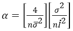
DN= “Digital Number (values within the moving window)”
K= “normalization constant”
 = “local weighted mean calculated by using Equation 2”
σ2 = “local variance”
 “image coefficient of variation value”
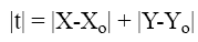
Where  refers to the modulus of difference from average
value in x direction and
refers to the modulus of difference from average
value in x direction and  refers to the difference of
modulus from average value in y direction n= moving kernel
size. In order to handle this issue, the Frost filter is modified by
taking the weighted local average of the degraded/corrupted
image. The existing filter is using the simple average [22,23]
while the correlation of pixels is very important that is why the
proposed adaptive weighted filter uses the weighted average of
the local window. The Weighted average of empirically chosen
5 × 5 Window is computed using Equation 2. Perceptibly, a
filter that considers the correlation with the adjacent pixels
inside the moving window would create the comparatively
better enhancement results.
refers to the difference of
modulus from average value in y direction n= moving kernel
size. In order to handle this issue, the Frost filter is modified by
taking the weighted local average of the degraded/corrupted
image. The existing filter is using the simple average [22,23]
while the correlation of pixels is very important that is why the
proposed adaptive weighted filter uses the weighted average of
the local window. The Weighted average of empirically chosen
5 × 5 Window is computed using Equation 2. Perceptibly, a
filter that considers the correlation with the adjacent pixels
inside the moving window would create the comparatively
better enhancement results.
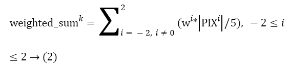
The proposed modified Adaptive Weighted Frost filter is hence a ratio based filter that uses weighted sum of values within N × N window in order to replace the central pixel. When the correlation with central and remaining pixels decreases, the weighting factor also decreases and so on. The modified Frost filter is elaborated in the Equation 2 with 5 × 5 window size:
The proposed weighted sum formula is presented in the Equation 2.
 (2)
(2)
Where
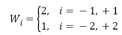
The proposed weighted window shown in the Figure 2 will be used to calculate the weighted mean.

Figure 2: Weighted window for weighted local mean.
If the lag distance of two pixels is less, then usually the gray scale values are closer to each other. So accordingly as presented in Figure 2, more weight is assigned to the pixels whose lag distance is 1 and less weight whose lag distance is 2 to create the correlation among adjacent pixels. Thus the high correlation of connected pixels is achieved which is very important here since the adjacent pixels contribute well in order to restore the central pixel of degraded image. Consequently, the resultant pixel value has been replaced by the weighted mean of filter window. That is why the proposed Adaptive Weighted Frost filter is better to the existing Frost and Wiener filter for quantum noise removal from mammograms.
Performance measure
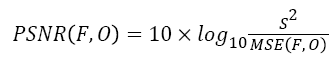
Peak signal-to-noise ratio (PSNR) is taken as a quantitative measurement in order to evaluate the performance of the proposed Adaptive Weighted Frost filter to remove the quantum from mammograms using the Equation 3 [19]. The PSNR value ranges from 30 to 50 db is considered acceptable which means that the visual quality of mammogram is improved.
 (3)
(3)
Where,
O denotes the “original mammogram”,
F represents the “restored mammogram”,
S refers to the value of maximum intensity (255) and
MSE is the Mean Square Error which is presented in Equation 4 [19].
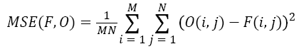 (4)
(4)
Results and Discussion
In order to test the proposed methodology, all the experiments were conducted using Mammographic Image Analysis Society (MIAS) - a benchmarked dataset [21]. The modified adaptive Frost filter successfully eliminate the noise from mammograms, the resultant mammogram is enhanced and noise free images which can be used for further processing like segmentation and classification.
Concerning the post-noise-removal visual quality of mammographic images, the proposed scheme was benchmarked with state-of-the-art and well-known techniques, which were proposed by Naveed et al. [19], and very famous filters, Frost filter [22] and Wiener filter [19,24], and the quantitative results are presented in Table 1, Figure 3a and Figure 3b. In order to remove noise, Naveed et al. proposed a combination of Frost filter, adaptive wiener filter and non-local mean filter.
| Image/Method | Proposed Method | Combination of Three Filters | Frost Filter | Wiener Filter |
|---|---|---|---|---|
| mdb001 | 34.6 | 28.4 | 27.9 | 26.9 |
| mdb005 | 36.3 | 33.2 | 32.1 | 28.7 |
| mdb007 | 37.8 | 33.5 | 30.9 | 31.1 |
| mdb010 | 39.1 | 31.4 | 31.3 | 26.9 |
| mdb025 | 34.7 | 28.7 | 28.5 | 27.6 |
| mdb041 | 34.1 | 31.2 | 27.3 | 29.1 |
| mdb064 | 35.5 | 30.6 | 25.1 | 31 |
| mdb081 | 31.5 | 29.1 | 27.4 | 24.9 |
| mdb099 | 37.9 | 31.8 | 31.5 | 25.2 |
| mdb101 | 39.8 | 29.7 | 28.5 | 23.8 |
| mdb110 | 36.3 | 30.7 | 27.8 | 29.1 |
| mdb118 | 37.6 | 31.5 | 29 | 23.7 |
| mdb130 | 35.3 | 28.4 | 26.7 | 25.7 |
| mdb143 | 40.5 | 32.7 | 29.3 | 28.9 |
| mdb162 | 37.6 | 29.9 | 24 | 27.5 |
| mdb178 | 35.8 | 32.3 | 31.7 | 25.6 |
| mdb199 | 35.4 | 30.6 | 26.5 | 27.1 |
| mdb206 | 38.4 | 31.5 | 29.4 | 31.1 |
| mdb219 | 42.9 | 33.5 | 27.3 | 23.2 |
| mdb244 | 32.9 | 31.4 | 28.1 | 27.4 |
| mdb275 | 36.1 | 30.6 | 26.9 | 25.8 |
| mdb313 | 33.4 | 29.8 | 31.5 | 21.9 |
| mdb320 | 42.4 | 28.9 | 27.5 | 26.4 |
| mdb322 | 44.7 | 31.6 | 28.4 | 28.7 |
Table 1: PSNR comparison of proposed technique with existing techniques.

Figure 3A: PSNR comparison of proposed technique with existing techniques.

Figure 3B: PSNR comparison of proposed technique with existing techniques.
Relatively, it is obvious from the overhead results that the proposed method scored higher PSNR values while comparing to its counterparts. This is the indication of high image quality and visually better results achieved by the proposed technique- PSNR above 30 db is considered good. In order to reaffirm the above outcomes, an additional experiment is conducted. This time, the noise is intentionally added to original mammographic images using Matlab Poisson noise function, and the results are shown in Figure 4. It is noticed that the Poisson noise has been removed successfully and the sharpness of the images are maintained; the images almost resembled their original.

Figure 4: Noise removal: (A-A2) Original image (B-B2) Noise added image and (C-C2) Filtered image.
The current study used a state-of-the-art preprocessing technique in order to enhance the quality of mammograms to removal quantum noise. The Adaptive Frost filter is modified and implemented to preprocess the mammograms for the detection of breast cancer. A quantitative measure Peak signalto- noise ratio is calculated to check the performance of proposed filter. The experimental results revealed that adaptive the proposed Adaptive Frost filter is the best to remove the noise from mammograms. It is quite clear from the results that the proposed technique achieved higher PSNR values as compared to other three existing techniques. It demonstrates that the quality of mammograms attained by the proposed technique is visually better and the PSNR more than 30 db is considered acceptable which shows that the noise is restored from mammograms and now they are fit for further processing.
Conclusion
The presented noise restoration technique by using modified Frost filter to improve the quality of mammographic images showed promising results. The results demonstrated that the restored mammograms showed low noise level (high PSNR) and better spatial resolution (resemblance to their originals and no artifacts are produced in mammogram images). The preprocessed mammographic images showed better qualitative and quantitative results which are helpful for further processing such as features extraction, micro-calcification detection, breast cancer diagnosis, classification, etc. The proper preprocessing steps also increase the efficiency of CAD (computer aided diagnosis) systems which are used by radiologists for abnormalities detection. The modified Frost filter produced better PSNR values, for example in mdb001, the PSNR values produced by modified Frost filter is 34.6 db, Naveed et al. is 28.4 db, Frost filter 27.9 db and Wiener filter is 26.9 db. The same trend is shown by all mammographic images which illustrates the supremacy of proposed modified filter for mammograms restoration.
Acknowledgements
The authors are thankful to the Deanship of Scientific Research, King Saud University Riyadh Saudi Arabia for funding through the Research Group Project no. RG-1437-037.
References
- Siegel R, Naishadham D, Jemal A. Cancer Statistics, 2013. CA: Cancer JClinicians 2013; 63: 11-30.
- Wang H, Li JB, Wu L,Gao H. Mammography visual enhancement in CAD-based breast cancer diagnosis. ClinImaging 2013; 37: 273-282.
- dos Santos Teixeira RF. Automatic Analysis of Mammography Images, Classification of Breast Density. Master Thesis, University of Porto 2013.
- Jemal A, Siegel R. Cancer statistics, 2010.CA:Cancer JClinicians 2010; 60: 277-300.
- Smith RA, Cokkinides V, Brawley OW. Cancer screening in the United States, 2012. CA: Cancer J Clinicians2012; 62: 129-142.
- Mandelblatt JS, Cronin KA, Bailey S, Berry DA, de Koning HJ, Draisma G, HuangH, Lee SJ, Munsell M, Plevritis SK. Effects of mammography screening under different screening schedules, model estimates of potential benefits and harms. AnnalInternal Medicine 2009; 151: 738-747.
- SundaramKM,SasikalaD, Rani PA. A Study On Preprocessing A Mammogram Image Using Adaptive Median Filter. Int J Innovative Res SciEngTechnol 2014; 3: 10333-10337.
- Liu B, Cheng HD, Huang J, Tian J, Tang X, Liu J. Fully automatic and segmentation-robust classification of breast tumors based on local texture analysis of ultrasound images. Pattern Recognition 2010; 43: 280-298.
- Serifovic-Trbalic A, Trbalic A, Demirovic D, Prljaca N,Cattin P. Classification of benign and malignant masses in breast mammograms. Info CommunTechnol Electronics Microelectronics (MIPRO) 2014; 2014: 228-233.
- Acharya RU, Ng EYK, Chang YH, Yang J, Kaw GJL. Computer-Based Identification of Breast Cancer Using Digitized Mammograms. J Medical Systems 2008; 32: 499-507.
- DheebaJ,SelviST. A Swarm Optimized Neural Network System for Classification of Microcalcification in Mammograms. J Medical Systems 2008; 36: 3051-3061.
- Zakeri FS, Behnam H, Ahmadinejad N. Classification of benign and malignant breast masses based on shape and texture features in sonography images. J Medical Systems 2012; 36: 1621-1627.
- Sampat MP, Markey MK. Computer-aided detection and diagnosis in mammography. Handbook of Image and Video Processing 2005; 2: 1195-1217.
- Mustra M,GrgicM. Robust automatic breast and pectoral muscle segmentation from scanned mammograms. Signal Processing 2013; 93: 2817-2827.
- Dos Santos RLC, da Costa VMA,Schiabel H. Mammography images restoration by quantum noise reduction and inverse MTF filtering. IEEE Brazilian SympComput Graphics Image Process (SIBGRAPI) 2009.
- Mencattini A, Salmeri M.Noise estimation in mammographic images for adaptive denoising. EFOMP European Conference on Medical Physics 2007.
- Elmore JG, Nakano CY, Koepsell TD, Desnick LM, D’Orsi CJ, Ransohoff DF. International variation in screening mammography interpretations in community-based programs. J Nat Cancer Institute 2003; 95: 1384-1393.
- Bhateja V, Verma A, Rastogi K, Malhotra C, Satapathy SC. Performance Improvement of Decision Median Filter for Suppression of Salt and Pepper Noise. Advances in Signal Processing and Intelligent Recognition Systems 2014, Springer: 287-297.
- Naveed N, Hussain A, Arfan JM, Choi TS. Quantum and impulse noise filtering from breast mammogram images. ComputMethods Programs Biomed 2012; 108: 1062-1069.
- Scholkmann F, Revol V, Kaufmann R, Baronowski H,Kottler C. A new method for fusion, denoising and enhancement of x-ray images retrieved from Talbot-Lau grating interferometry. Physics Med Biol2014; 59: 1425-1440.
- Suckling J, Parker J.The Mammographic Images Analysis Society digital mammogram database.ExerptaMedicaInt Congress Series 1994; 1069: 375-378.
- Frost VS, Stiles JA, Shanmugan KS,Holtzman JC. A model for radar images and its application to adaptive digital filtering of multiplicative noise. IEEE Transactions on Pattern Analysis and Machine Intelligence 1982.
- Huang Y,Genderen JLV. Evaluation of Several Speckle Filtering Techniques for ERS-1 & 2 Imagery. Int Arch Photogrammetric Remote Sensing 1996; 31: 164-169.
- Lim JS. Two-dimensional Signal and Image Processing, Prentice Hall, Englewood Cliffs, NJ, 1990, p. 548, equations 9.44-9.46.