ISSN: 0970-938X (Print) | 0976-1683 (Electronic)
Biomedical Research
An International Journal of Medical Sciences
Research Article - Biomedical Research (2018) Volume 29, Issue 14
Multimodal image fusion using an evolutionary based algorithm for brain tumor detection
1Department of Computer Science, Sathyabama Institute of Science and Technology, Chennai, India
2Department of Computer Science, Sri Venkateswara College of Engineering, Sriperumpudur, India
- *Corresponding Author:
- Jany Shabu SL
Department of Computer Science
Sathyabama Institute of Science and Technology
Chennai, India
Accepted date: July 01, 2018
DOI: 10.4066/biomedicalresearch.29-18-799
Visit for more related articles at Biomedical ResearchMedicinal image fusion is an unpredictable procedure. Joining and combining data from at least two images from various sources or modalities into a solitary image is known as image fusion. This kind of joining of various data's from two unique modalities is done to build the indicative ability. In this work, genetic algorithm (GA) based image combination is performed with the assistance of continuous medicinal images. Hereditary algorithm is an evolutionary procedure. For combination we utilize two critical GA administrators called hybrid and change. The proposed system is performed on two arrangements of magnetic resonance imaging (MRI) and, computed tomography (CT) images. The execution of the proposed system is assessed with various quality measurements.
Keywords
Fusion, Genetic algorithm, Brain tumor detection.
Introduction
Image combination is a procedure of image preparing in which converging of at least two images into a solitary composite image, that coordinates the data of both source images with no contortion or clamor. Multi-modular restorative image combination is a novel strategy utilized by doctors to perceive the sores or anatomical changes. To break down images of various modalities intertwining images is another and sparkling region of research [1]. This territory of research is noticeable as a result of the requests in clinical applications. The zone of biomedical image handling is a skilled zone of research from most recent two decades [2]. Therapeutic imaging process is sub isolated as useful and auxiliary data related preparing. Magnetic resonance imaging (MRI) and computed tomography (CT) manages high-determination images by methods for basic while methodology like positron emission tomography (PET) and single-photon emission computed tomography (SPECT) images gives us the practical information's. The objective of image combination is to acquire the data at every pixel area of the information images without harming the pixel relations of that image. Along these lines protected data portrays the genuine substance of that image. It additionally improves the melded image for a superior diagnosis. The problem in image combination is that it if the information images are undermined one because of commotions at that point however compelling the combination might be yet the data retrieved from the fused image will be most exceedingly awful only. So before performing combination we must be careful that the different methodology images which are chosen for combination is preprocessed and clamor free. Clamors are mainly because of movement ancient rarity's as well as because of old machines utilized for imaging. In old imaging machines, uncalled for centering of radiation or inappropriate constriction are major problems. To keep away from this sort of commotions we need to apply a suitable channel which disposes of the above said clamors such has salt and pepper clamor and Gaussian or drive clamors.
A novel strategy for DWT-GA based combination calculation for intertwining MRI or PET images with CT images is proposed. Related work clarifies the writing work and the proposed technique is clarified in methodology, results and talks are given with quantitative analysis followed by conclusion.
Related Work
Singh et al. proposed a complex wavelet change for image combination. They have utilized most extreme choice run for combination [3]; The comes about were contrasted and LWT, MWT, SWT, NSCT, DTCWT and PCA strategies. Most extreme choice control is connected for various arrangements of multimodal images. It is inferred that the nature of combined image increments, as the level of decay increases.
Ellmauthaler et al. proposed a combination technique which includes undecimated wavelet change [4]. In this procedure the image is disintegrated into two consecutive sifting tasks. Ghastly factorization is utilized as a part of the separating procedure. Best outcomes are gotten acquired when UWT with 2 tap Haar channel bank is used. It is additionally noticed that is reasoned that Haar channel presents antiquities and disintegrate the execution of image combination process at the recreation part.
Lu et al. proposed the utilization of most extreme nearby vitality strategy and the result were compared with wavelets [5]. The coefficients of the diverse methodology were obtained. Low-recurrence and high recurrence coefficients are then chosen by most extreme neighbourhood vitality and entirety changed Laplacian strategies. The yield image is delivered by taking converse wavelet change. The outcomes show that the most extreme nearby vitality approach is ideal.
Liu et al. proposed an agreeable based combination mode, where SWT and NSCT are actualized [6]. The source image is decayed by SWT and NSCT. The NSCT coefficients and the SWT coefficients and NSCT coefficients are joined to create the melded image.
Upadhyay et al. proposed a method where the images treated with SWT initially and then the global textural features are obtained from the gray level co-occurrence matrix [7-10].
Proposed Methodology
As combination assumes a noteworthy part in image combination, to intertwine image after pre-handling we have utilized Genetic Algorithm (GA). Genetic algorithm (GA) is utilized to produce the combination rules and there by playing out the combination procedure. The basic schematic portrayal of the proposed framework is given underneath in Figure 1. The proposed system is clarified in the beneath segment.

Figure 1: Proposed methodology block diagram.
The initial step is to obtain distinctive methodology images like PET and CT images as information. Next the info images are changed over to dark scale and resized. Subsequent stage is to improve the images, for this we have utilized the imadjust order accessible in MATLAB. We have utilized median channel for commotion expulsion. Middle channel is great in expelling salt and pepper commotions, which is generally present in medicinal images. This is because of the movement antiquities or because of the old gadgets utilized for checking purposes. Execution measurements like PSNR, contrast, entropy and so forth were utilized.
PSNR signifies the level of clamor accessible in the image. It is the esteem which tells whether our yield melded image has ruined or not. In the event that so the PSNR esteem will be less and the MSE esteem will be vast. MSE esteem ought to be constantly less and the PSNR esteem ought to be constantly high.
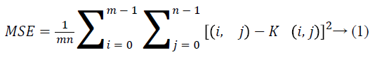

Equations 1 and 2 gives the MSE and PSNR values.
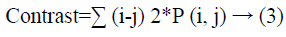
GLCM highlights, for example, energy, contrast, and homogeneity were additionally established to assess our process. Energy discloses to us how much data is available in the image. Contrast restores the force data about the pixel with its neighbour. The condition for computing contrast is given beneath.
Homogeneity tells the measure of closeness of elements present in the gray level concurrence matrix (GLCM).
Proposed System
The optimization technique is used to create something better than the existing system. The genetic algorithm (GA) is an optimization technique that is used to improve the fusion of medical images like MRI and CT to highlight the region of interest like tumor portion after resultant image of the fusion. The genetic algorithm (GA) requires the input images as any format with noise free and high quality images for further image fusion. The image fusion using genetic algorithm is an optimization technique only for the high quality that the image should high adaptive contrast and brightness without any kind of noise. This quality is mandatory for image fusion using GA that is useful for finding out the tumor portion on the image. The genetic algorithm is implemented from the concept of the theory by Charles Darwin. It stated that GA implements Darwinian survival of the fitness on the population of creature. In all production original position of persons are makes and only the fittest persons will get a possibility to donate to the next generations. The algorithm is designed based on genetic optimization rule to reduce the impact of requirements changes on the development image fusion. With a purpose to recognize its full capability, there are tools and systems wished for the responsibilities intrinsic to the evolutionary algorithm. On this proposed system, we implement genetic algorithm is used to build software program development and upkeep work as genetic set of rules have robustness and genetic algorithm is commonly used to generate top mark solutions to optimization and seek troubles by counting on simulated operators which includes mutation, crossover and selection.
The curvelet transform is applied on the input images MRI and CT to convert the spatial domain information to frequency domain information. The concept of curvelet transform is initially decomposing the input image into sub-bands. It represents the image in different scales and projections during the transformation. Curvelet assumes anisotropic scaling while wavelet assumes isotropic projection. The anisotropic projection and scaling are more accurate than wavelets. Curvelt obeys algebraic scaling law, i.e., width=length^2. Curvelt transform into curves if they are scaled. The below is the flow diagram of genetic optimization rule (Figure 2).

Figure 2: The flow chart of genetic algorithm.
The genetic algorithm optimization technique
Algorithm for a GO is as follows:
Initialize (population)
Evaluate (population)
While (stopping condition not satisfied) do
{
Selection (population)
Crossover (population)
Mutate (population)
Evaluate (population)
}
Fitness function
Fitness function and variables perform that should be maximized or decreased is termed as Fitness perform or value perform related to the fitness perform is termed as fitness value or value. Supported this criterion it will be determined whether or not a selected individual would contribute to future generation or not. The variables or parameters on that the fitness performs depend are on the array and is referred as chromosome.
Consider, the fitness function F holds on variable V1, V2, V3… Vn. Thus an individual chromosome will be written as follows,
Chromosome=(V1, V2, V3, ,… Vn)
Cost=f (Chromosome)=f (V1, V2, …, Vn)
Here we take the entropy of the resultant fusion image as the cost function and variable considered with this cost function are weights of the input images. As defined above the technique is derived on images MRI and CT are Wmri and Wct be the weights of individual images respectively then the cost function and chromosome is as given below.
Cost function=entropy (Wmri × MRI+Wct × CT)
Where, MRI-->Magnetic Resonance Image
CT-->Computer Tomography
Chromosome=(Wmri, Wct)
Initial population
The initial population will be randomly chosen. If the region of solution is known, then the principles within that region of solution can also be selected as initial population. In the medical image fusion technique, the weights of the input images can be in the range of [0, 1] a matrix size Np × 2 have random values between 0 to 1 is chosen and it is considered as to be initial population. Where, Np is the total number of chromosomes available in the initial population generation. Every row of this matrix is selected as chromosome and its corresponding fitness value (entropy of fused image) is estimated and then the matrix is rearranged in descending manner depends on the fitness values.
Selection and mating
The individuals generated as initial population only the fittest are selected to supply to the next generation of genetic optimization rule. In the matrix estimated only the top ‘Nk’ elements are kept and the remaining elements are eliminated. The performance of the proposed algorithm is depends on the genetic operations, basically, on the type of the crossover methodology. The crossover is the basic operation performed as a part of genetic optimization rule. This operation is reproduction upon which the genetic algorithm is working. During the evaluation of genetic algorithm, if the estimated chromosome is same, then some of the crossover operations have unsuccessful. This failure will create offspring that are different their patents chromosomes. In this procedure more than one possibility of the present generation is considered and is created to create the new generation solutions. The present generation solutions taken for crossover are called parents; the new solutions produced are called as children.
Mutation
The mutation process is used to alter one or more generation values in a chromosome from its first state. This genetic operation is used to maintain genetic diversity and to eliminate quick convergence of genetic algorithm. If the algorithm converges very quickly then it will lead wrong mutation then to false maximum.
Tumor segmentation
After applying image fusion using genetic optimization rule, the input images MRI and CT are fused together. The main aim of image fusion is that the highlighting the useful information from different types of images are achieved after applying image fusion technique. The fuzzy based image segmentation is applied to segment the region of interest (ROI) to separate the tumor portion from the fused image. The adaptive fuzzy c means clustering rule is applied to segment the region of interest from the fused image. The adaptive fuzzy c means clustering technique will cluster the different gradient intensity portion of the fused image. The high gradient intensity portion pixels are the tumor pixels that are separated by applying the thresholding technique [11-13].
Results and Discussions
It is important to break down the proposed technique from both subjective and target focuses. Here the execution of the proposed work is contrasted and five arrangements of continuous restorative images got from various Scans in Chennai, Tamil Nadu-India. For the proposed philosophy five arrangements of combination conspire is connected.
The accompanying Figures from 3 to 6 shows the info CT and MR images and the melded yield images which are connected on the proposed calculation.
Debauchee’s wavelet is utilized for choosing the combination rules. Here in this examination we have utilized the base run the show. Diverse levels of decay give distinctive levels of combination. The deterioration level ought not to be low or high. The level decides the level of defilement or clamor information misfortune in the image. Figures 3 and 4 are dim scale CT and MRI images.

Figure 3: I/P gray scale CT image.

Figure 4: I/P gray scale MRI image.
Figures 5 and 6 are for median filtered CT and MRI Images. Figure 7 shown is the final fused output image. This image has good subjective enhancement.

Figure 5: Filtered CT image.

Figure 6: Filtered MRI image.

Figure 7: Fused image.
Figures 8 and 9 are the decomposed output CT and MRI images. Based on the minimum rule the low values of CT are fused with the low level decomposition of the MRI image.

Figure 8: Wavelet decomposition of MRI.

Figure 9: Decomposition of CT image.
The numerical values for the quantitative measurements such have PSNR, Contrast, Entropy is set of CT and MRI images are given in Table 1.
| MSE | PSNR | Entropy | Contrast | Homogeneity | |
|---|---|---|---|---|---|
| SET1 | 14851 | 6.4132 | 11.7865 | 0 | 1 |
| SET2 | 11826 | 7.4023 | 12.0226 | 0 | 1 |
| SET3 | 18168 | 8.6945 | 12.864 | 0 | 1 |
| SET4 | 12413 | 7.5624 | 12.4568 | 0 | 1 |
Table 1: Quantitative measures of the proposed algorithm.
Conclusion
A novel pixel based image combination technique joined with wavelet and Genetic calculation is proposed in this exploration article. From the perception of the outcomes and the qualities for entropy, mean square error, peak signal to noise ratio (PSNR), correlation, and homogeneity obviously demonstrates that the intertwined image is of better quality. Hereditary calculation is a developmental based calculation which helps in improvement as well as gives a versatile calculation composition to image combination calculations. It helps in the choice of best combination rules. As to build the proficiency of our proposed calculation we have used second request middle separating. In view of the level of separating the precision of image combination depends. Traverse and mutation administrators are utilized as GA administrators in this exploration work.
References
- Lei W, Bin L, Lian-Fang T. Multi-modal medical image fusion using the inter-scale and intra-scale dependencies between image shift-invariant shearlet coefficients. Elsevier 2014; 19: 20-28.
- Sabalan D, Hassan G. MRI and PET image fusion by combining IHS and retina-inspired models. Elsevier 2010; 11: 114-123.
- Rajiv S, Ashish K. Fusion of multimodal medical images using Daubechies complex wavelet transforms-A multi-resolution approach. Elsevier 2014; 19: 49-60.
- Andreas E, Carla LP, Da Silva AB. Multiscale image fusion using the undecimated wavelet transform with spectral factorization and non-orthogonal filter banks. IEEE Transactions on Image Processing 2013; 22: 1005-1017.
- Huimin L, Lifeng Z, Seiichi S. Maximum local energy: An effective approach for multisensor image fusion in beyond wavelet transform domain. Elsevier 2012; 64: 996-1003.
- Yi L, Guanzhong L. Cooperative fusion of stationary wavelet transform and non-subsampled contourlet for multifocus images. IEEE Second International Symposium on Computational Intelligence and Design 2009; 1: 314-317.
- Manoj DC, Abhay BU. Fusion of local and global features using stationary wavelet transform for efficient content based image retrieval. IEEE Students Conference 2014; 1-6.
- Po-Whei H, Cheng-IC, Phen-Lan L. PET and MRI brain image fusion using wavelet transform with structural information adjustment and spectral information patching. IEEE Conference 2014; 1-4.
- Tapasmini S. Removal from satellite images using auto associative neural network and stationary wavelet transform. IEEE 2008; 100-105.
- Hailiang S, Min F. Multi-focus color image fusion based on SWT and HIS. IEEE Conference 2007; 461-465.
- Boulerbah C, Takieddine S, Aichouche B. Unsupervised change detection from multitemporal multichannel SAR images based on stationary wavelet transforms. IEEE Conference 2013; 1-4.
- Jany Shabu SL, Jaya Kumar C. Multimodal image fusion and bee colony optimization for brain tumor detection. ARPN J Eng Appl Sci 2018; 13: 1819-6608.
- Xiaoqing Z, Yongguo Z, Yanjun P. Research on multi-mode medical image fusion algorithm based on wavelet transform and the edge characteristics of images. IEEE Conference 2009; 1-4.