ISSN: 0970-938X (Print) | 0976-1683 (Electronic)
Biomedical Research
An International Journal of Medical Sciences
- Biomedical Research (2013) Volume 24, Issue 1
Incidence of Supratrochlear foramen of Humerus in North Indian Population
Department of Anatomy, King George’s Medical University, Lucknow-226003 (U.P.), India
- *Corresponding Author:
- Rakesh Kumar Diwan
Department of Anatomy
King George’s Medical University
Lucknow-226003 (U.P.), India
Accepted date: November 26 2012
A thin translucent plate of bone usually separates coronoid and olecranon fossa of humerus. In some humerii this plate is perforated and gives rise to an aperture, known as supratrochlear foramen. The present study was conducted on 1776 dried humerii (Right-905, left-871) of which 1352 belong to males and 424 to females. The sex determination of humerus was determined by using Vernier sliding calipers by measuring the maximum length, circumference of midshaft, vertical and transverse diameters of head and maximum medial epicondylar breadth of humerus to document the incidence of supratrochlear foramen in North Indian population. The transverse and vertical diameter of the supratrochlear foramen was measured by using digital Vernier calipers.. The supratrochlear foramen was found in 24.10% of humerii. The incidence was more common on left side (28.13%) as compared to right (20.22%). Oval, round and triangular shapes of the supratrochlear foramen were observed among which the oval shape was found to be maximum. The knowledge of higher incidence of supratrochlear foramen in Indians will be helpful to orthopaedic surgeons and radiologists in treating and interpreting the pathology in this area respectively.
Keywords
Humerus, Supratrochlear foramen, translucent septum
Introduction
The coronoid and olecranon fossa of humerus are usually separated by a thin translucent plate of bone. In certain bones this plate is sometimes perforated by a foramen, called the supratrochlear foramen (STF) or septal aperture. STF in humerii was first described by Meckel in 1825 [1]. Hardlicka observed that the perforation is more frequent in higher primates other than man [2]. Incidence of STF varies from 6% to 60% in different races [3]. The anatomical knowledge of STF may be beneficial for anthropologists, orthopaedic surgeons and radiologists in day-to-day clinical practice. The present study describes the incidence and different shapes of supratrochlear foramen in the humerii of North Indian population.
Material and Methods
A total of 1776 (Right 905, Left-871) dried adult humerii of which 1352 belongs to male and 424 to female of North Indian origin were collected from the osteology lab of the Department of Anatomy, King George’s Medical University, U.P. Lucknow. The observed bones were free from any pathology. The transverse and vertical diameter of the supratrochlear foramen was measured by using digital Vernier calipers. The shape of the foramen was also seen.
Results
The supratrochlear foramen was present in 428 (24.1%) of humerii in the present study. The total incidence of STF was higher in left sided humerus both in males and females. The incidence was 28.13% on left side while it was 20.22% on right side. In males, it was seen in 26.76% cases on left side while 21.45% cases on right side. In females, STF was present in 31.89% humerus on left side and 15.63% on right side (Table 1).
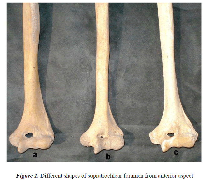
The shape of supratrochlear foramen also varied in different humerii. Three types of shape was observed, i.e. oval, round and triangular (Fig 1). Among them majority showed oval foramen i.e. 83.06% (n=152) on right side and 82.04% (n=201) on left side. The incidence of round foramen was seen in 15.30% (n=28) on right side and 15.10% (n=37) on left side. Only 10 humerus showed triangular foramen i.e.1.63% (n=3) on right side and 2.85% (n=7) on left side (Table 2). The size of different shapes of STF were measured and was found that the average transverse and vertical diameter in the round shaped STF was 0.28 mm on right side and 0.23 mm on left side. The mean vertical diameter of oval shaped STF was 3.6 mm on right side and 3.8 mm on left side while the mean transverse diameter was on both right and left side was 5.5 mm. The mean height in triangular shaped STF was 3.1 mm on right side and 3.06 mm on left side while the length of the base was 4.73 mm on right side and 4.22 mm on left side. In 1348 humerii, there was no septal aperture between coronoid and olecranon fossa. In these bones, either translucent or opaque septum was observed. Translucent septum was present in 91.13% (n=658) while opaque septum was seen in 8.86% (n=64) of right sided humerii. The incidence of translucent septum was 79.39% (n=497) while opaque septum was 20.60% (n=129) in left sided humerii.
Discussion
The presence of supratrochlear foramen is considered to be an atavistic character, as it is frequently found in primates [2]. In Indian population, the incidence is different in different regions. In Eastern Indians it is 27.4% [4], in Central Indians 32% [1], in South Indians 28% [3], while in North Indians it is 27.56% [5]. In the present study of North Indian population the incidence is 24.10% which is in line with the findings of Singh & Singh and suggest that the occurrence is slightly on the lower side as compared to other Indians. In Americans it is 6.9 % [6], in Egyptians 7.9% [7], in Ainus 8.8% [8], and in Japanese the incidence is 18.1% [8] (Table 3).
The majority of bones that had no foramen showed translucency of septum (91.13% on right side and 79.39% on left side)), but Singhal et al observed translucent septum in 66% humerii of South Indian origin [3]. In their study the opaque septum was present in 6% bones which is lower as compared to North Indians observed in the present study, i.e 8.86% on right side and 20.60% on left side. Majority of the STF were oval in shape (82.47%) which strengthens the previous reports by Singhal et al and Nayak et al followed by round and triangular shape [3,10].
The higher incidence of the STF in the Indian population requires special attention during intramedullary humeral nailing procedures in the distal portion of humerus. In the present era, intramedullary fixation has become a popular method of treatment, especially in traumatic injuries and pathological fractures due to its advantages. Therefore, The frequency of supratrochlear foramen in our study was found to be higher in females and more common on left side (Table 1). This finding was similar to previous reports of Singh & Singh who also observed that STF was present more in females (38%) than in males (21%) and more frequently on the left side (31%) than on the right side (24%) [5]. Comparative sidedness of STF in different human races shows that in majority of population it is more frequent on left side (Table 4). Mall suggested that the higher occurrence of STF in females may be due to inward curvature of the female elbow angle [4]. According to Hirsh (1927) the thin plate of bone between the olecranon and coronoid fossa is always present until the age of seven years after which the bony septum occasionally becomes absorbed to form the STF [9].
The presence of such foramen should be kept in mind before proceeding for surgical intervention. Prior anatomical knowledge about the presence of STF may check erroneous interpretation of X-rays by radiologists. De Wilde et al opined that STF is an area which is relatively radiolucent, is commonly seen as a type of ‘pseudo- lesion’ in any X-ray of the upper limb, and this can be mistaken for an osteolytic or cystic lesion [11].
References
- Meckel JH (1825) cited by Kate BR, Dubey PN. A note on the septal apertures in the humerus of Central Indians. Eastern Anthropologist 1970; 33: 105-110.
- Hardlicka A. The humerus septal apertures. Anthroplogie 1932; 10:34-96.
- Singhal S, Rao V. Supratrochlear foramen of the humerus. Anat Sci Int 2007; 82: 105-107.
- Mall (1905) cited by Chatterjee KP. The incidence of perforation of olecranon fossa in the humerus among Indians. Eastern Anthropologist 1968; 21:270-284.
- Singh, Singh SP. A study of the supratrochlear foramen in the humerus of North Indians. J Anat Soc India 1972; 21: 52-56.
- Benfer, McKern T. The correlation of bone robusticity with the perforation of the coronoid-olecranon septum of humerus of man. Am J Phys Anthropol 1966; 24: 247-252.
- Ozturk A, Kutlu C, Bayraktar B, Zafer ARI, Sahinoglu K. The supratrochlear foramen in the humerus (Anatomical study). st Tp Fak. Mecmuas 2000; 63: 72-76.
- Akabori E. Septal apertures in the humerus in Japanese, Ainu and Koreans. Am J Phys Anthropol 1934;18:395-400.
- Hirsh SI (1927), cited in Morton SH, Crysler WE. Osteochondritis dissecans of the supratrochlear septum. J Bone Joint Surg 1945; 27-A: 12-24.
- Nayak SR, Das S, Krishnamurthy A, Prabhu LV, Potu BK. Supratrochlear foramen of the humerus: An anatomico-radiological study with clinical implications. Upsala Journal of Medical Sciences. 2009; 114: 90-94.
- De Wilde V, De Maeseneer M, Lenchik L, Van Roy P, Beeckman P, Osteaux M. Normal osseous variants presenting as cystic or lucent areas on radiography and CT imaging: a pictorial overview. Eur J Radiol 2004; 51:77-84.


