ISSN: 0970-938X (Print) | 0976-1683 (Electronic)

Biomedical Research

An International Journal of Medical Sciences

- Biomedical Research (2016) Volume 27, Issue 1

In vitro genotoxicity evaluation of tungsten (VI) oxide nanopowder using human lymphocytes.

Bugra G. Akbaba1*, Hasan Turkez2, Erdal Sonmez3, Ugur Akbaba4, Elanur Aydin5, Abdulgani Tatar6, Guven Turgut7, Salim Cerig5
1Bioengineering Department, Faculty of Engineering and Architecture, Kafkas University, Kars, Turkey
2Department of Molecular Biology and Genetics, Faculty of Science, Erzurum Technical University, Erzurum, Turkey
3Graduate School of Natural and Applied Sciences, Department of Nanoscience & Nanoengineering, Advanced Materials Research Laboratory, Atatürk University, Erzurum, Turkey
4Department of Elementary Mathematics Education, Education Faculty, Kafkas University, Kars, Turkey
5Atatürk University, Faculty of Science, Department of Biology, 25240, Erzurum, Turkey
6Medical Genetics Department, Faculty of Medicine, Ataturk University, Erzurum, Turkey
7Department of Physics. K. K Education Faculty, Atatürk University, Erzurum, Turkey
Corresponding Author: Dr. Giray Bugra Akbaba, Kafkas University, Faculty of Engineering and Architecture, Bioengineering Department, 36100, Kars/Turkey.
Accepted December 24, 2015
Visit for more related articles at Biomedical Research

Abstract

Tungsten (VI) oxide (or tungsten trioxide) (WO3, <100 nm particle size) nanoparticles (NPs) are used for many purposes including production of electrochromic windows, or smart windows, x-ray screen and gas sensors in everyday life. However, their carcinogenicity and genotoxicity have not been sufficiently evaluated. Therefore, the genotoxic potential of WO3 nanoparticle was examined in cultured human lymphocytes by the use of the micronucleus (MN) test and the comet (SCGE) assay. Freshly isolated human lymphocytes were exposed to WO3 nanoparticle at concentrations ranging from 0 to 500 μM for 72 hours at 37°C. Our results indicated that 400 and 500 μM of WO3 nanoparticle treatment caused slight increases of the MN frequencies in cultured human lymphocytes. Likewise, WO3 nanoparticle (at concentrations above 200 μM) led to increases of DNA damage (estimated with the comet assay) in human lymphocytes. The observed alterations in the MN and the comet assay parameters revealed that WO3 nanoparticles have genotoxic potential and could pose environmental and human health risk.

Keywords

Genotoxicity, Health risk, Human lymphocytes, Nanoparticle, Tungsten trioxide

Introduction

Nanoparticles (NPs) are identified as particles with diameters under 100 nm, are unique in that their electronic, chemical, and physical properties enable many promising technical and medicinal applications [1,2]. Thanks to their unique features, NPs have been the focus of much research such as in industrial applications, environmental toxicity studies and human health impacts. Various industrial NPs are made from titanium oxide, silver, gold, cadmium selenite, other carbon NPs, cerium oxide and hydroxyapatite NPs [3-6]. Together with the fast development of nanotechnology today, NPs are used for various biomedical applications such as targeted delivery/ imaging, hyperthermia, cell therapy and stem cell tracking [7-14]. In recent years, many efforts were made to investigate the toxicity of micro sized natural and man-made mutagens to human life and the ability of therapeutic substances to reduce the toxicity of these chemicals [15-20]. But the toxic effects of NPs were not fully detailed except for some inorganic and organic NPs. In fact, the most recent report indicated that there was a lack of systematic assessment of the DNA damaging and carcinogenic potential of NPs in spite of their extensive use in nanotechnological applications. People are exposed to NPs from various sources and in many pathways, including inhalation, dermal absorption, eye contact and oral ingestion [21,22]. Therefore, the evaluation of NPs toxicity has become very important for public health and the environment [23-25].
Tungsten trioxide contains oxygen and the transition metal tungsten. It is gained as an intermediate in the recovery of tungsten from its minerals. To produce tungsten products tungsten ores are treated with alkalis. Tungsten trioxide can be prepared in several different ways. Scheelite (CaWO4) is allowed to react with HCl to produce tungstic acid, which decomposes to WO3 and water at high temperatures. Another common way to synthesize WO3 is by calcination of ammonium paratungstate (APT) under oxidizing conditions. There are many applications of the WO3 in everyday life. It is used in industry to manufacture tungstates for fireproofing fabrics, for x-ray screen, in gas sensors, automobile glass and as a pigment in ceramics and paints because of its rich yellow color [26-30]. In recent years, WO3 has been employed in biomedical applications as an endovascular coil, endovascular catheter and bone cement [31-33]. Although tungsten had been considered a relatively inert and toxicologically safe material, recent research findings have raised concerns about possible deleterious health effects after acute and chronic exposure to this metal [34, 35]. It was reported that soluble tungsten compounds were absorbed after oral exposure both in humans and in laboratory rats. It has been shown that the embedded tungsten alloy pellets caused metastatic tumors in rats. Tungsten was found to accumulate in several organs and/or tissues such as kidneys, liver, ovaries, prostate, pancreas, lung, heart, muscle, spleen and bone following a single oral dose [36]. In addition, a previous report indicated the potential for tungsten alloy-induced immunotoxicity [37]. The genotoxic potential of tungsten and tungsten compounds has not been extensively assessed [38]. Considering the latest information, the mutagenic potential of WO3 nanopowder has not been accurately perused. Thus, in this paper, we thoroughly investigate the cytotoxic and genotoxic potentials of WO3 nanoparticles in human lymphocytes culture by using the micronucleus (MN) test and the comet (SCGE) assay.
Materials and Methods

Synthesis of tungsten trioxide nanoparticles

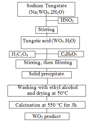
Metal oxide based semiconductors such as SnO2, ZnO, TiO2, CuO and WO3 etc. have been used in many application areas [39]. Among these, WO3 is one of the most valuable materials for electrochromic devices, information displays, smart windows and rechargeable lithium batteries [40]. WO3 as transitional metal oxide not only has reversible electrochromism property and special catalysis property [41]; at one time because of its big surface area, WO3 can be used excellent solar absorb material and contact material: but also WO3 belonging to an n-type semiconductor has excellent gas sensing property [42]. In the recent years, nanopowder WO3 materials have gained much attention due to its surface to volume ratio, which is much greater than that of coarse-grained materials [43]. There is a wide range of techniques for preparation of the powders such as the sol-gel process [44], the micro emulsion method [42], the inert gas condensation method and the chemical vapor condensation process [45]. Among these techniques, the sol-gel technique is attractive due to its easy manipulation of the samples, simplicity, safety, low cost [46], and easy control of chemical components [47]. The sol-gel method involves the dispersion of metallic salts in solutions. The sol is later ?solidified? through stages of stiffening and polymerization to give a gel (gelation) [48]. The gel so obtained is thoroughly washed with distilled water or alcohol, filtered, dried and finally heated to high temperatures to obtain the required material [49]. Therefore, in present study, WO3 nanopowders were prepared via the sol-gel process. The experimental construction is shown in Figure 1. Firstly, nitric acid (HNO3) solution was added drop by drop to sodium tungstate (Na2WO4.2H2O), so tungstic acid deposit was formed. Oxalic acid (H2C2O4) and citric acid (C6H8O7) were used as complex forming agents in the sol solution. The precipitate obtained from this solution was washed several times with absolute ethyl alcohol and then dried at 50°C. In this manner yellow precipitates were formed and WO3 powder was produced with calcination at 550°C for 3h.
image
Figure 1: Schematic diagrams of steps involved in obtaining WO3 powders

Lymphocyte cultures

Typically, two or three donors have been used in toxicity studies [18,19]. In this study, four donors were used to increase the statistical reliability. Human peripheral blood samples were drawn from healthy volunteers (age = 30 years), by venipuncture in heparinized tubes. Leukocytes (lymphocytes +monocytes) were isolated on a Ficoll-Paque gradient, washed with Phosphate Buffered Saline (PBS) and resuspended in Ham?s F10 medium containing 15% foetal calf serum (FCS). Lymphocytes were stimulated to divide by 2% phytohaemagglutinin (PHA). Cultures were set up at a concentration of 0.5 Ã

References

  1. Pan Y, Leifert A, Ruau D, Neuss S, Bornemann J, et al. Gold nanoparticles of diameter 1.4 nm trigger necrosis by oxidative stress and mitochondrial damage. Small 2009; 5: 2067-2076.
  2. Warheit DB, Hoke RA, Finlay C, Donner EM, Reed KL, et al. Development of a base set of toxicity tests using ultrafine TiO2 particles as a component of nanoparticle risk management. Toxicol Lett 2007; 171: 99-110.
  3. Park EJ, Choi J, Park YK, Park K. Oxidative stress induced by cerium oxide nanoparticles in cultured BEAS-2B cells. Toxicology 2008; 245:90-100.
  4. Cockburn A, Bradford R, Buck N, Constable A, Edwards G, et al. Approaches to the safety assessment of engineered nanomaterials (ENM) in food. Food Chem Toxicol 2012; 50: 2224-2242.
  5. Turkez H, Sonmez E, Turkez O, Mokhtar YI, Stefano AD, et al. The Risk Evaluation of Tungsten Oxide Nanoparticles in Cultured Rat Liver Cells for Its Safe Applications in Nanotechnology. Braz Arch Biol Technol 2014; 57: 532-541.
  6. Di Stefano A, Sozio P, Iannitelli A, Cerasa LS. New drug delivery strategies for improved Parkinson’s disease therapy. Expert Opin Drug Deliv 2009; 57: 532-541.
  7. Stephan MT, Moon JJ, Um SH, Bershteyn A, Irvine DJ. Therapeutic cell engineering with surface-conjugated synthetic nanoparticles. Nat Med 2010; 16: 1035-1041.
  8. Mahmoudi M, Hosseinkhani H, Hosseinkhani M, Boutry S, Simchi A, et al. Magnetic resonance imaging tracking of stem cells in vivo using iron oxide nanoparticles as a tool for the advancement of clinical regenerative medicine. Chem Rev 2010; 111: 253-280.
  9. Lee JH, Jang J, Choi J, Moon SH, Noh S, et al. Exchange-coupled magnetic nanoparticles for efficient heat induction. Nat Nanotechnol 2011; 6: 418-422.
  10. Irvine DJ. Drug delivery: One nanoparticle, one kill. Nat Mater 2011; 10: 342-343.
  11. Di Stefano A, Iannitelli A, Laserra S, Sozio P. Drug delivery strategies for Alzheimer’s disease treatment. Expert Opin Drug Deliv 2011; 8: 581-603.
  12. Chauhan VP, Stylianopoulos T, Martin JD, Popovic Z, Chen O, et al. Normalization of tumour blood vessels improves the delivery of nanomedicines in a size-dependent manner. Nat Nanotechnol 2012; 7: 383-388.
  13. Noh S, Na W, Jang J, Lee J-H, Lee EJ, et al. Nanoscale magnetism control via surface and exchange anisotropy for optimized ferrimagnetic hysteresis. Nano Lett 2012; 12: 3716-3721.
  14. Geyikoglu F, Turkez H, Keles SM. The role of fruit juices in the prevention of aluminum sulphate toxicity in human blood in vitro. Fresen Environ Bull 2005; 14: 878-883.
  15. Geyikoglu F, Türkez H. Protective effect of sodium selenite on genotoxicity to human whole blood cultures induced by aflatoxin B1. Braz Arch Biol Technol 2005; 48: 905-910.
  16. Geyikoglu F, Turkez H. Boron compounds reduce vanadium tetraoxide genotoxicity in human lymphocytes. Environ Toxicol Phar 2008; 26: 342-347.
  17. Turkez H, Tatar A, Hacimuftuoglu A, Ozdemir E. Boric acid as a protector against paclitaxel genotoxicity. Acta Biochim Pol 2010; 57: 95.
  18. Turkez H, Togar B. The genotoxic and oxidative damage potential of olanzapine in vitro. Toxicol Ind Health 2010; 26: 583-588.
  19. Yah CS. The toxicity of gold nanoparticles in relation to their physiochemical properties. Biomed Res 2013; 24: 400-413.
  20. Mostofi R, Wang B, Haghighat F, Bahloul A, Jaime L. Performance of mechanical filters and respirators for capturing nanoparticles-limitations and future direction. Ind Health 2010; 48: 296-304.
  21. Win-Shwe TT, Fujimaki H. Nanoparticles and neurotoxicity. Int J Mol Sci 2011; 12: 6267-6280.
  22. Oberdörster G, Oberdörster E, Oberdörster J: Nanotoxicology. An emerging discipline evolving from studies of ultrafine particles. Environ Health Persp 2005; 823-839.
  23. Li SQ, Zhu RR, Zhu H, Xue M, Sun XY, et al. Nanotoxicity of TiO2 nanoparticles to erythrocyte in vitro. Food Chem Toxicol 2008; 46: 3626-3631.
  24. Liu Z, Liu S, Ren G, Zhang T, Yang Z. Nano-CuO inhibited voltage-gated sodium current of hippocampal CA1 neurons via reactive oxygen species but independent from G-proteins pathway. J Appl Toxicol 2011; 31: 439-445.
  25. Lassner E, Schubert WD. Tungsten: Properties, Chemistry, Technology of the Element, Alloys, and Chemical Compounds. Kluwer Academic, USA; 1999.
  26. Lee WJ, Fang YK, Ho JJ, Hsieh WT, Ting SF, et al. Effects of surface porosity on tungsten trioxide (WO3) films’ electrochromic performance. J Electron Material 2000; 29: 183-187.
  27. Williams DE, Aliwell SR, Pratt KF, Caruana DJ, Jones RL, et al. Modelling the response of a tungsten oxide semiconductor as a gas sensor for the measurement of ozone. Meas Sci Technol 2002; 13: 923.
  28. Patnaik P. Handbook of Inorganic Chemical Compounds McGraw-Hill handbooks. McGraw-Hill Professional 2002; 1086.
  29. The Merck Index. Tungsten trioxide. 2006; Vol. 14.
  30. Butler TJ, Jackson RW, Robson JY, Owen RJ, Delves HT, et al. In vivo degradation of tungsten embolisation coils. Br J Radiol 2000; 73: 601-603.
  31. Peuster M, Kaese V, Wuensch G, Wuebbolt P, Niemeyer M, et al. Dissolution of tungsten coils leads to device failure after transcatheter embolisation of pathologic vessels. Heart 2001; 85: 703-704.
  32. Hierholzer J, Depriester C, Fuchs H, Venz S, Maier-Hauff K, et al. Rofo-Fortschr Rontg 2002; 174: 328-334.
  33. Guandalini GS, Zhang L, Fornero E, Centeno JA, Mokashi VP, et al. Tissue distribution of tungsten in mice following oral exposure to sodium tungstate. Chem Res Toxicol 2011; 24: 488-493.
  34. Witten ML, Sheppard PR, Witten BL. Tungsten toxicity. Chem-Biol Interact 2012; 196: 87-88.
  35. McInturf SM, Bekkedal MY-V, Wilfong E, Arfsten D, Gunasekar PG, et al. Neurobehavioral effects of sodium tungstate exposure on rats and their progeny. Neurotoxicol Teratol 2008; 30: 455-461.
  36. Kalinich JF, Emond CA, Dalton TK, Mog SR, Coleman GD, et al. Embedded weapons-grade tungsten alloy shrapnel rapidly induces metastatic high-grade rhabdomyosarcomas in F344 rats. Environ Health Persp 2005; 113: 729-734.
  37. Keith LS, Moffett DB, Rosemond ZA, Wohlers DW. ATSDR evaluation of health effects of tungsten and relevance to public health. Toxicol Ind Health 2007; 23: 347-387.
  38. Singh MP, Singh H, Singh O, Kohli N, Singh RC. Preparation and characterization of nanocrystalline WO3 powder based highly sensitive acetone sensor. Indian J Phys 2012; 86: 357-361.
  39. Li YB, Bando Y, Golberg D, Kurashima K. WO3 nanorods/nanobelts synthesized via physical vapor deposition process. Chem Phys Lett 2003; 367: 214-218.
  40. Pandey NK, Tiwari K, Roy A. Ag Doped Nanomaterials as Relative Humidity Sensor. IEEE Sens J 2011; 11: 2911-2918.
  41. Hou CJ, Diao XZ, Tang YK, Huo DQ, Wei LF. Preparation and Characterization of WO3 Nano-powder with Microemulsion Method. 2nd IEEE International Nanoelectronics Conference 2008; 362-365.
  42. Siegel RW. Nanostructured materials-mind over matter. Nanostruct Mater 1994; 4: 121-138.
  43. Kanan SM, Tripp CP. Synthesis, FTIR studies and sensor properties of WO3 powders. Curr Opin Solid St M 2007; 11: 19-27.
  44. Maruyama T, Arai S. Electrochromic properties of tungsten trioxide thin films prepared by chemical vapor deposition. J Electrochem Soc 1994, 141: 1021-1024.
  45. Ghodsi FE, Absalan H. Comparative study of ZnO thin films prepared by different sol-gel route. Acta Phys Pol A 2010; 118: 659-664.
  46. Ilican S, Caglar Y, Caglar M. Preparation and characterization of ZnO thin films deposited by sol-gel spin coating method. J Optoelectron Adv M 2008; 10: 2578-2583.
  47. Poulter KF, Pryde JA. Chemisorption of hydrogen on rhenium. J Optoelectron Adv M 1968; 1: 169.
  48. Han SD, Campet G, Huang SY, Shastry MCR, Portier J, et al. A new method for the preparation of fine-grained SnO2 and WO3 powders: influence of the crystallite size on the electrochemical insertion of Li+ in SnO2 and WO3 electrodes. Act Passive Electron Compon 1995; 18: 39-51.
  49. Sontakke YA, Fulzele RR. Cytogenetic study on genotoxicity of antitumor-antibiotic Mitomycin C. Biomed Res 2009; 20: 40-44.
  50. Singh NP, McCoy MT, Tice RR, Schneider EL. A simple technique for quantitation of low levels of DNA damage in individual cells. Exp Cell Res 1988; 175: 184-191.
  51. Karaman A, Kadi M, Kara F. Sister chromatid exchange and micronucleus studies in patients with Behcet’s disease. J Cutan Pathol 2009; 36: 831-837.
  52. Erol A. Systemic DNA damage response and metabolic syndrome as a premalignant state. Curr Mol Med 2010; 10: 321-334.
  53. Turkez H, Geyikoglu F, Mokhtar YI, Togar B. Eicosapentaenoic acid protects against 2, 3, 7, 8-tetrachlorodibenzo-p-dioxin-induced hepatic toxicity in cultured rat hepatocytes. Cytotechnology 2012; 64: 15-25.
  54. McKelvey-Martin VJ, Green MHL, Schmezer P, Pool-Zobel BL, De Meo MP, et al. The single cell gel electrophoresis assay (comet assay): a European review. Mutat Res Fund Mol M 1993; 288: 47-63.
  55. Turkez H, Cakmak B, Celik K. Evaluation of the potential in vivo genotoxicity of tungsten (VI) oxide nanopowder for human health. In Key Engineering Materials. Volume 543. Trans Tech Publ 2013; 89-92.
  56. Hasegawa G, Shimonaka M, Ishihara Y. Differential genotoxicity of chemical properties and particle size of rare metal and metal oxide nanoparticles. J Appl Toxicol 2012; 32: 72-80.
  57. Kühnel D, Scheffler K, Wellner P, Meißner T, Potthoff A, et al. Comparative evaluation of particle properties, formation of reactive oxygen species and genotoxic potential of tungsten carbide based nanoparticles in vitro. J Hazard Mater 2012; 227: 418-426.
  58. Papageorgiou I, Brown C, Schins R, Singh S, Newson R, et al. The effect of nano-and micron-sized particles of cobalt?chromium alloy on human fibroblasts in vitro. Biomaterials 2007; 28: 2946-2958.
  59. Lovric J, Cho SJ, Winnik FM, Maysinger D. Unmodified cadmium telluride quantum dots induce reactive oxygen species formation leading to multiple organelle damage and cell death. Chem Biol 2005; 12: 1227-1234.
  60. Jin Y, Kannan S, Wu M, Zhao JX. Toxicity of luminescent silica nanoparticles to living cells. Chem Res Toxicol 2007; 20: 1126-1133.
  61. Dubertret B, Skourides P, Norris DJ, Noireaux V, Brivanlou AH, et al. In vivo imaging of quantum dots encapsulated in phospholipid micelles. Science 2002; 298: 1759-1762.
  62. Guilbert C, Kelly AD, Petruccelli LA, Lemaire M, Mann KK. Exposure to tungsten induces DNA damage and apoptosis in developing B lymphocytes. Leukemia 2011; 25: 1900-1904.
  63. Oberdörster G, Maynard A, Donaldson K, Castranova V, Fitzpatrick J, et al. ILSI Research Foundation/Risk Science Institute Nanomaterial Toxicity Screening Working Group. Principles for characterizing the potential human health effects from exposure to nanomaterials: Elements of a screening strategy. Part Fibre Toxicol 2005; 2: 8.
  64.