ISSN: 0970-938X (Print) | 0976-1683 (Electronic)
Biomedical Research
An International Journal of Medical Sciences
Research Article - Biomedical Research (2017) Volume 28, Issue 6
Foot pressure distribution indicates the osteoporosis and the fall risk of older persons
1Department of Endocrinology, Affiliated Hospital of Qiingdao University, PR China
2Department of Endocrinology, the 401 Hospital of PLA, PR China
3Department of Endocrinology, Oingdao Municipal Hospital, PR China
4Licang District Central Hospital of Qingdao, PR China
5Weifang Hospital of Traditional Chinese Medicine, PR China
- *Corresponding Author:
- Nailong Yang
Department of Endocrinology
Affiliated Hospital of Qiingdao University, PR China
Accepted date: November 04, 2016
Falls is one of the most important problems for older persons, which usually causes the elder disability. The indication and prevention of the falls are one of the major concerns in the elder cares. Several risk factors and risk assessment tools including foot pressure for falls were investigated for the hospital inpatients. However, none of them is ready for popularizing in non-patients lived in common communities. Here, we intend to investigate the correlations between the foot pressure characters and the risk of falls in older persons (age ≥ 60 years old). 218 healthy elders without falling history lived in common communities were involved in this study and their pressure distribution of both feet during static standing was measured by Footscan USB2 system. Ultrasonic was used to measure the bone density of calcaneus to evaluate the osteoporosis. We found out that the individuals with osteoporosis showed abnormal local pressure increase compared to non-osteoporosis individuals. The falling occurrence was followed during the two years after the measurements. We found out that the individuals with abnormal pressure distribution, including significant pressure increase in the area of toes (without hallux), third and fifth metatarse, and the lateral arch (LA), and the asymmetrical pressure distribution between the two feet, showed higher fall risk than other individuals. The individuals with osteoporosis showed higher risk of falls. Our study suggested foot pressure measurement combined with osteoporosis screening as a powerful procedure to evaluation the fall risk in older non-patients lived in common communities.
Keywords
Foot pressure distribution, Fall risk, Elder, Osteoporosis.
Introduction
Population ageing, which is a phenomenon that occurs when the median age of a country or region increases due to rising life expectancy and/or declining fertility rates. It is one of the most concern problems around the world [1-3]. Facing population ageing, fall prevention is a crucial public health concern, because falls occurred in more than one third of people aged over 65 each year, resulting in injury, disabled functioning, and mortality [4-6]. After falling, about 5% elders will undergo bone fracture; about 5-11% elders will have severe injury, which is one of the main lethal causes of the elders. The medical cost after the falling is about 6% of the total for the elders.
Falling is a multifactorial result relevant to environments, individuals’ physiological, pathological, and mental conditions. It was recognized that the factors leads to falling included the instability of gait, the disturbance of consciousness, the urinary incontinence or frequent urination, falling history, and administration of tranquilizer or hypnotics, etc. [4]. Targeted interventions and multifactorial fall risk assessment, environmental inspection along with hazard-reduction programs, and exercise programs are recommended to reduce the incidence and adverse consequences from falls [7-9]. However, recent investigations about fall prevention suggests that systematic fall risk assessment and intervention may only reduce a modest amount of the fall risk or fall incidence rates or may even be non-contributing [4].
Dozens of methods and scales were invented to evaluate the fall risk [10], including three most investigated scales - STRATIFY, Hendrich II fall risk model, and Morse fall scale [11]. However, most of these methods or scales were investigated for evaluating the fall risk of hospitalized elders. The investigations about evaluating the falling risk for elders live in the common communities are not conclusive. Here, by measuring the pressure distribution of pelma using Foot scan USB2 system combined with evaluating osteoporosis by calcaneus ultrasound, we found out that both abnormal pressure distribution of pelma and osteoporosis can be served as indicators for fall risk in non-patients in community.
Materials and Methods
Subjects
Persons above the age of than 60 years old lived in the common communities (Qingdao, Shandong Province, China) were recruited as subjects in this study. 218 elders (105 males and 113 females) with no falling history were included. The demographic characteristics, including age, height, weight, and foot length were recorded and showed in Table 1. This study was approved by the Medical Ethics Committee of Affiliated Hospital of Qiingdao University (2015-12-30) and is in accordance with the recent principles of the Declaration of Helsinki. All patients received and signed the informed consent and were free to withdraw from the study at any time.
| Gender | Male | Female |
|---|---|---|
| Number | 105 | 113 |
| Age (years) | 68.10 ± 5.27 | 71.72 ± 7.25 |
| Height (cm) | 164.28 ± 3.22 | 157.96 ± 4.32 |
| Weight (kg) | 66.32 ± 8.22 | 61.07 ± 13.22 |
| Foot length (cm) | 24.83 ± 0.82 | 22.24 ± 0.24 |
| Osteoporosis (%) | 59.05 | 88.50 |
| Mean ± SD for age, height, weight and foot length. | ||
Table 1. Demographic characteristics.
Foot pressure distribution measurement
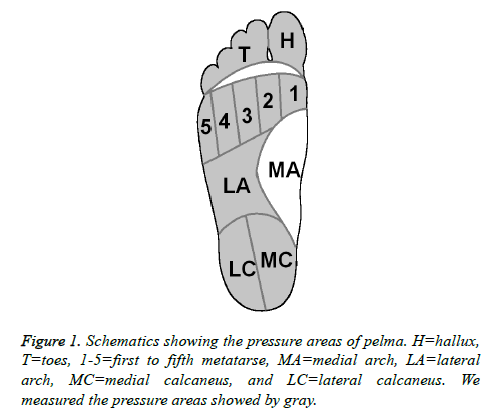
Footscan USB2 system (RSscan International, Belgium) was used to measure the pressure distribution of pelma as described before [12]. Pressures of ten areas, including hallux (H), toes (T), first to fifth metatarse (1-5), medial arch (MA), lateral arch (LA), medial calcaneus (MC), and lateral calcaneus (LC), were recorded (Figure 1) when the subjects were static standing on the system.
Bone density measurement of calcaneus
Calcaneus density was measured using ultrasound as described before [12,13]. Measurement of calcaneal ultrasound was undertaken using a Sahara clinical bone sonometer (Hologic Inc., Bedford, MA) without prior knowledge of osteoporosis diagnosis and treatment status.
Asymmetrical pressure between left and right feet
The asymmetry of the two feet was evaluated using an asymmetric score. Briefly, the asymmetric score is an integral reflecting the asymmetry of maximum pressures in the ten areas of the two feet. The differences of the maximum pressures between the two feet were evaluated using the Pearson's chi-squared test. If P<0.05, it gives a factor of one in this area, otherwise it gives a factor of zero. The final asymmetric factor is an integral of the ten areas, which is a value from 0-10 and reflect the asymmetrical pressure between left and right feet.
Statistics
The data sets were presented as mean values ± standard deviations (SD). The statistical differences of the mean values were evaluated using the student t-test. The statistical differences of the falling occurrence rate were evaluated using Pearson's chi-squared test. The statistical differences between left and right feet in each individual were evaluated using the Pearson's chi-squared test. P<0.05 was considered as significant difference.
Results
Individuals with osteoporosis showed abnormal pelma local pressure and higher risk of falling compared to non-osteoporosis individuals
We found out that in our overall older 105 males and 113 females, 62 males (59.05%) and 100 females (88.5%) were identified as osteoporosis (Table 1). The individuals with osteoporosis showed a significantly different pelma pressure distribution (Table 2) compared to the non-osteoporosis individuals. Particularly, there’s a significant pressure decrease in the area of the first metatarse (from ~13 kPa to ~9 kPa) and a significant pressure increase in the area of the fourth and fifth metatarses (from ~6 kPa and ~6 kPa to ~17 kPa and ~10 kPa, respectively) for the both feet. Notably, there’s also some significant increase of decrease in some areas for just one foot (left or right), which results more asymmetrical pressure distribution between the two feet in the osteoporosis individuals (nine of the ten areas showed significant difference in the pressure between the two feet) compared to the nonosteoporosis individuals (three of the ten areas showed significant difference in the pressure between the two feet). After the two years of our measurement, 17 of the 218 individuals underwent falling during these two years. Interestingly, we found that 16 of them have and only 1 of them has not diagnosed as osteoporosis before. Thus, 16 in the overall 162 osteoporosis individuals underwent falling, which gave a falling occurrence rate of 9.88%. This is significantly higher than the one of the non-osteoporosis individuals (1.79%, 1 in 56).
| Areas | Osteoporosis (n=162) | non-osteoporosis (n=56) | ||
|---|---|---|---|---|
| Left foot | Right foot | Left foot | Right foot | |
| H | 8.9 ± 2.2* | 12.4 ± 1.9# | 11.8 ± 2.5 | 12.3 ± 3.1 |
| T | 9.3 ± 2.1 | 3.1 ± 2.4#* | 9.6 ± 3.0 | 4.5 ± 2.8# |
| 1 | 9.7 ± 4.3* | 9.1 ± 3.2* | 13.4 ± 2.8 | 12.6 ± 3.9 |
| 2 | 11.4 ± 4.3* | 16.7 ± 3.8# | 18.4 ± 1.9 | 17.6 ± 4.5 |
| 3 | 17.5 ± 3.7 | 16.1 ± 2.8#* | 18.1 ± 2.6 | 24.8 ± 6.2# |
| 4 | 16.3 ± 4.1* | 18.9 ± 3.7#* | 5.7 ± 3.4 | 6.5 ± 2.8 |
| 5 | 8.4 ± 1.1* | 13.9 ± 2.3#* | 6.1 ± 3.4 | 5.1 ± 2.1 |
| LA | 6.2 ± 2.5* | 10.3 ± 4.2# | 12.6 ± 3.2 | 9.2 ± 3.9# |
| MC | 15.8 ± 3.2 | 13.1 ± 2.8#* | 17.2 ± 4.4 | 16.8 ± 4.8 |
| LC | 17.4 ± 2.7* | 12.9 ± 1.9#* | 16.1 ± 3.1 | 15.3 ± 3.4 |
| *P<0.05 when compared with non-osteoporosis group using student t-test; #P<0.05 when compared with left foot in the same group using student t-test. | ||||
Table 2. Maximum pressure of different areas on pelma between osteoporosis and non-osteoporosis individuals (kPa).
Pelma local pressure indicate the falling risk
To assess whether pelma local pressure can indicate the falling risk, we divided the 218 individuals to two groups by the pressure of the ten pelma areas, respectively. The pressure medians of the ten pelma areas were used as the cut-offs to split the groups, respectively (so we have ten kinds of splitting, Table 3). Interestingly, we found out that the pressure increase in the area of toes (without hallux), third and fifth metatarse, and the lateral arch (LA) showed higher risk of falling. Higher pressure in the area of toes increases the falling occurrence rate from 2.75% to 12.84%. Higher pressure in the area of third metatarse increases the falling occurrence rate from 3.67% to 11.93%. Higher pressure in the area of fifth metatarse increases the falling occurrence rate from 4.59% to 11.01%. Higher pressure in the area of LA increases the falling occurrence rate from 0.92% to 14.68%.
| Areas (Median/kPa) | Falling occurrence rate (%) | |
|---|---|---|
| Maximum pressure<median(n=109) | Maximum pressure>median(n=109) | |
| H (11.0) | 7.34 | 8.26 |
| T (6.4) | 2.75 | 12.84* |
| 1 (10.3) | 8.26 | 7.34 |
| 2 (15.1) | 7.34 | 8.26 |
| 3 (18.0) | 3.67 | 11.93* |
| 4 (14.6) | 6.42 | 9.17 |
| 5 (9.7) | 4.59 | 11.01* |
| LA (8.9) | 0.92 | 14.68* |
| MC (15.1) | 7.34 | 8.26 |
| LC (15.3) | 8.26 | 7.34 |
| *P<0.05 when compared with maximum pressure<median group using Pearson's chi-squared test. | ||
Table 3. Comparison of the falling occurrence in the following two years between different groups.
Furthermore, we assessed the correlations of the pelma pressure asymmetry and the falling risk. We introduced an asymmetric score (0-10), which based on the asymmetry integrity of the ten pelma pressure areas, to evaluate the pelma pressure asymmetry of each individual. From Table 4, we can see there are two populations in the entire individual based on the asymmetric score, one population with low asymmetric score (~3) and one population with high asymmetric score (~8), which is consistent with the finding that the osteoporosis increase the asymmetry. We found out that only two individuals, which underwent falling in the past two years, have a low asymmetric score (0-5). However, 15 individuals, which underwent falling in the past two years, have a high asymmetric score (6-10). The falling occurrence rate in the low asymmetric score individuals is 3.13% (2 in 64), which is much lower than the falling occurrence rate in the high asymmetric score individuals (9.74, 15 in 154).
| Asymmetric score | Total individuals | Falling occurrence | Percentage (%) |
|---|---|---|---|
| 0 | 0 | 0 | 0 |
| 1 | 2 | 0 | 0 |
| 2 | 14 | 0 | 0 |
| 3 | 30 | 1 | 3.333333333 |
| 4 | 10 | 0 | 0 |
| 5 | 8 | 1 | 12.5 |
| 6 | 10 | 0 | 0 |
| 7 | 21 | 2 | 9.523809524 |
| 8 | 79 | 7 | 8.860759494 |
| 9 | 41 | 4 | 9.756097561 |
| 10 | 3 | 2 | 66.66666667 |
Table 4. Falling occurrence in the groups with different asymmetric score.
Discussion
These results indicated that osteoporosis can change the pressure distribution on the pelma and increase the asymmetry, which may affect the balance of the elders and lead to falling. Facing aging societies, fall prevention is a critical public health concern [14]. The methods to evaluating the fall risk for elders are very important. An efficiency method must be fast and valid. Here, we introduce a method to evaluating the fall risk by measure the pressure distribution on pelma combined with osteoporosis diagnosis. We find out that the individuals with osteoporosis have higher risk of fall, which may due to the abnormal pelma pressure distribution after osteoporosis [15,16]. When we only see the pressure distribution on pelma, we found out that the pressure increase in the area of toes (without hallux), third and fifth metatarse, and the LA indicate higher risk of fall. Furthermore, the pelma pressure asymmetry is correlated with fall risk, which makes sense that the unbalance and the instability of gait is one of the main causes of falls. This study present a convenient method for the screening in common community, which can effectively indicate the fall risk and will greatly help elders prevent falls. In conclusion, the pelma local pressure can indicate the falling risk by pressure increase in specific areas and pelma pressure asymmetry. However, the extension of this foundation still needs further research with big samples and multi-center.
References
- Briggs AM, Cross MJ, Hoy DG, Sanchez-Riera L, Blyth FM, Woolf AD. Musculoskeletal Health Conditions Represent a Global Threat to Healthy Aging: A Report for the 2015 World Health Organization World Report on Ageing and Health. Gerontologist 2016; 56: S243-255.
- Smith BD, Smith GL, Hurria A, Hortobagyi GN, Buchholz TA. Future of cancer incidence in the United States: burdens upon an aging, changing nation. J Clin Oncol 2009; 27: 2758-2765.
- Ortman JM, Velkoff VA, Hogan H. An Aging Nation: The Older Population in the United States. US CENSUS BUREAU 2014: 1-28.
- Lee HC, Chang KC, Tsauo JY, Hung JW, Huang YC, Lin SI. Effects of a multifactorial fall prevention program on fall incidence and physical function in community-dwelling older adults with risk of falls. Arch Physical Med Rehab 2013; 94: 606-615.
- Holroyd-Leduc JM. Review: exercise interventions reduce falls in elderly people living in the community. Evidence-Based Med 2009; 14: 176.
- Rubenstein LZ. Falls in older people: epidemiology, risk factors and strategies for prevention. Age Ageing 2006; 35: ii37-ii41.
- Campbell AJ, Robertson MC. Implementation of multifactorial interventions for fall and fracture prevention. Age Ageing 2006; 35: ii60-ii64.
- Rose DJ, Hernandez D. The role of exercise in fall prevention for older adults. Clin Geriatric Med 2010; 26: 607-631.
- Sherrington C, Whitney JC, Lord SR, Herbert RD, Cumming RG, Close JC. Effective exercise for the prevention of falls: a systematic review and meta-analysis. J Am Geriatrics Soc 2008; 56: 2234-2243.
- Oliver D, Daly F, Martin FC, McMurdo ME. Risk factors and risk assessment tools for falls in hospital in-patients: a systematic review. Age Ageing 2004; 33: 122-130.
- Ryan-Wenger NA, Kimchi-Woods J, Erbaugh MA, LaFollette L, Lathrop J. Challenges and conundrums in the validation of Pediatric Fall Risk Assessment tools. Pediatric Nurs 2012; 38: 159-167.
- Hessert MJ, Vyas M, Leach J, Hu K, Lipsitz LA, Novak V. Foot pressure distribution during walking in young and old adults. BMC Geriatrics 2005; 5: 8.
- Chin KY, Ima-Nirwana S. Calcaneal quantitative ultrasound as a determinant of bone health status: what properties of bone does it reflect? Int J Med Sci 2013; 10: 1778-1783.
- Li F, Harmer P, Fitzgerald K. Implementing an Evidence-Based Fall Prevention Intervention in Community Senior Centers. Am J Public Health 2016; 106: 2026-2031.
- Werner RA, Gell N, Hartigan A, Wiggermann N, Keyserling WM. Risk factors for foot and ankle disorders among assembly plant workers. Am J Indust Med 2010; 53: 1233-1239.
- Menz HB, Gill TK, Taylor AW, Hill CL. Predictors of podiatry utilisation in Australia: the north west Adelaide health study. J Foot Ankle Res 2008; 1: 1.
