ISSN: 0970-938X (Print) | 0976-1683 (Electronic)
Biomedical Research
An International Journal of Medical Sciences
- Biomedical Research (2016) Volume 27, Issue 3
Evaluating the thermal imaging system in detecting certain types of breast tissue masses.
1Department of Cellular and Molecular Biology, Semnan University, Semnan, Iran
2Department of Biomedical Engineering, Hakim Sabzevari University, Sabzevar, Iran
3Visual Communication Lab, Gwangju Institute of Science and Technology (GIST) University in Gwangju, South Korea
- *Corresponding Author:
- Hossein Ghayoumi Zadeh
Department of Electrical Engineering
Hakim Sabzevari University
Iran
Accepted date: February 24, 2016
The temperature of human body is indicative of critical medical information concerning the whole body. Abnormal rise in total and regional body temperature is a natural symptom in diagnosing many types of diseases. Thermal imaging (Thermography) utilizes infrared beams, which are fast, non-invasive, and non-contact. Generally, the output images by this technique are flexible and useful to monitor the temperature of the human body. The purpose of this study is to determine the diagnostic value of the breast tissue diseases by the help of thermography. In this paper, we used the non-contact infrared camera INFREC R500 for evaluating the capabilities of thermography. This study was conducted on 60 patients suspected of breast tissue disease, which were referred to Imam Khomeini Imaging Center. The overall information was obtained from multiple sources including a predefined medical questionnaire, the performed clinical examinations, diagnostic results obtained from ultrasound images, clinical biopsies and thermography. All of these inputs were analyzed by the respective experts of breast cancer. Medical analysis indicated that the use of Thermography as well as the asymmetry technique is useful in identifying hypo echoic and cystic masses. It should be noted that the patient should not suffer from breast discharge. The Accuracy of the asymmetry identification technique for identifying hypo echoic and cystic masses is 91/89% and 92/30%.In the same manner, the accuracy of identifying the exact location is 61/53% and 75%. This method is also effective in identifying heterogeneous, fibro-adenoma, and Intra-ductal masses. But it is unable to identify Iso-echo and calcified mass. According to the results of the investigation, Thermography is useful in the initial screening and supplementation of diagnostic procedures due to its safety (its non-ionizing radiation property which acts as everyday immersed heat), low cost and the exact recognition of diseases of the breast tissue.
Keywords
Thermography images, Biopsy, Breast cancer.
Introduction
Breast cancer is common in women and it is one of the leading causes of cancer death in women all over the world [1]. The most comprehensive statistical data on the prevalence of this disease has been established by the Center of diseases Control and Prevention. This data shows a dramatic increase in breast cancer during the last 50 years in the United States and after 30 years, a sudden increase occur in the rate of breast cancer, in addition to the short steady state that occurs between the ages of 45 and 50 years [2]. Furthermore, the World Health Organization (WHO) has estimated that in 2030, the rate of cancer in the world will get to 27 million people [3]. The most common type of cancer in women is breast cancer, and the second or the third and most common malignancy is in the developing countries [4]. There are several diagnostic methods for the detection of breast cancer. Early diagnosis is important for successful treatment. Due to the direct relationship between the delay of the early detection of the cancer and death resulting from it, the method that has characteristics such as being available, reliable and non-invasive is of great importance. Therefore, use of additional diagnostic techniques such as thermography can be of great benefit for patients [5]. Abnormal thermal patterns can be detected easily by thermal imaging. Generally, the findings of temperature measurement in comparison with other clinical findings are considered as a possible way to evaluate correlation. Although it is nonspecific method, sometimes it depends on the severity the background and the surrounding environment, but there are several reasons that have led thermal imaging achieve wide acceptance in the medical community. First of all, thermal imaging is a remote, non-contact and non-invasive method [6].The Imaging Time is very limited; therefore it is possible to monitor a large area from population simultaneously. Rendition of the Thermogram colors is easy and fast. Additionally, this method can record only natural radiation from the surface of the skin. And there is not any evidence from harmful rays. Therefore, it is suitable for a long term and repeated use.Finally; thermal imaging is a real-time method, which can monitor dynamic changes of temperature. The Radiation Potential for human dark skin is more or less equal to the constant value of 0.01 + 0.98 between wavelength 2 and 14 micrometers [7]. Therefore, in this area of wavelength, human skin acts as a black physical body, and due to its high absorption coefficient of 2/5 to 1/3mm at wavelengths between 2/2 and 5 micrometers, thermal radiation results from the outer surface of the skin [8]. Asymmetry Techniques are among the basic principles and methods for analyzing thermal images. Authors in [4] suggested that they used from an approach based on image segmentation by adopting image processing techniques such as Hough transform for the analysis of asymmetry. Analysis of asymmetry was based on the changes of temperature, skewness and kurtosis. Recently, according to the conducted studies in other countries it has been shown that Thermography imaging system gives the correct answer in the diagnosis of health or disease, by taking appropriate diagnostic and correct answers. And it would be appropriate office along with other methods as a complement approach [9-11].Studies based on existing technologies can be analyzed in both pre and post-2005. In the years before 2005, the 5 study, reported the accuracy of thermography as a diagnostic method in 9887 Lady with signs, symptoms or abnormal mammogram [12-16]. Average median or average age was 51 years during the study. The average age of the patients were 51 years old in this study. Diagnostic studies have been carried out before the screening time. The average rate qualities of these studies were mainly poor or fair. The main factor that reduces the quality of thermography in comparison to other methods is the lack of specialized knowledge required for perceiving the results of thermography and by other methods we mean CBE, mammography and biopsy. The average sensitivity of thermography itself in five previous diagnostic studies (ref? 1-5?) was 59% (range 25-97). When weak cases with less than the poor quality rate were deleted the average sensitivity decrease to 31% (range 25-47%) [12,17]. The highest sensitivity (95% and 97%) [14,15] was obtained in the test that patients with suspicious mammograms had been chosen. In these studies, the two term specificity/sensitivity exploited that when the sensitivity of the thermography is remarkably high (unlike most of the articles and unlike the estimated average) the specificity is very low (12% and 14%) [14,15]. These statistics indicate that thermography even in clinical trials with large sample sizes, has limited ability to discriminate between cancer and existence of changes. The five diagnostic Articles also report results for CBE in direct comparison with thermography, the mean sensitivity of CBE was 61% (range 51-86) [17-21]. In three of these five articles CBE is more sensitive than thermography [17,19,21]. Two papers reported Thermography sensitivity based on Tumors size [19,22]. These results have shown less sensitivity in tumor T1 (26-37%) compared with large tumors T2-4(over 82%). The average positive- false rate of thermography in diagnostic was 29% (range 8-86%). The average positive- false rate in both screening methods (average 31%) and diagnostic procedures (29%) in averages indicate that thermography will have a positive- false rate of about one times in every 3 patient. According to the approach and ability of Thermography, it can be used in to assess the effect of hormone receptor status (both estrogen and progesterone) in breast cancer [23]. This study has been done with the cooperation of 75 women with a mean age of 64 years, which had advanced breast cancer proven through biopsy. The output thermography results have been evaluated by the system, and the maximum, minimum and temperature deviation of the tumor areas have been detected. Also the all affected breast with tumor, healthy breast and breast with tumor regions have been identified. Thermography findings were compared with various status of the hormone receptor. Studies on hormone receptors showed the same results as thermography; the cancer tissue has a higher temperature than the healthy tissue. The study also demonstrated that among patients with different hormonal receptor status (positive or negative receptors), there is no significant statistical difference. However, these studies have shown that negative hormone receptor tumors are warmer tumors, show more invasive behavior, lose control of the endocrine glands and have a shorter life. Tumors with positive hormone receptor are cooler and show less invasive behavior. This information was not perceivable by thermography. Based on this study from all the patients having advanced breast cancer, 77% had positive receptor and 23% had negative receptor of estrogen. Also 60% had positive receptor and 40% had negative receptor of progesterone. By offering new models of thermal cameras and the use of modern technology, the accuracy in identification of tumors has increased the aid for diagnosis. Such as the three-dimensional thermal imaging method that has been evaluated in [24] where the obtained values of sensitivity and specificity in this article are 90.9% and 72.5%. In the experiment performed on 92 patients suffering from cancer, Biopsy testing was implemented and the obtained Value of sensitivity was 97% [14]. In a survey conducted in 2010, which was performed on 50 patients, the value of sensitivity was 78% and value of specificity was 75% [13]. In a study conducted on 49 patients with cancer, the value of sensitivity was 97%, and the value of specificity was 99%. The results of this study show that breast cancer screening by means of thermography is a very useful tool [25]. Notwithstanding the relative achievement of mammography, there is a need for ongoing research to increase the sensitivity of breast cancer recognition, especially younger women. Albeit mammography is currently considered to be the “gold standard” technology for the diagnosis of breast tumors the performance of this approach is less in younger women relates the hardness of imaging compact breast tissue and film interpretation. The main objective of the present study is to determine the usability and functionality of thermography system to identify some of the breast tissue disease compared with the ultrasound system.
Results
In this study, after receiving and recording the patient's history, the results of the clinical examination along with the ultrasound and biopsy stereotypes and images of thermograph were examined separately by the respective specialists. After recording the results, by considering the clinical signs, the capabilities of thermography techniques were compared in identifying whether the case was healthy or not. In general, 60 patients were studied, the mean age of which was 44/9 years, the youngest one was 21 years old and the oldest was 73. According to the conducted calculations, the standard deviation is 11.56 and Lconf and uoconf are equal to -0.2011 and 0.2011 respectively. As shown in Figure 1, after thermal imaging, the breast areas are initially separated symmetrically from left and right.

Figure 1. Analysis of thermal asymmetry in two thermal images with cancerous masses in the breast.
Then, the maximum and minimum and the average temperature are evaluated in these two areas. If the result doesn’t show significant difference, it is indicative of a non mass in the breast area (thermal symmetry). As shown in figure 1, the difference of one to two degrees Celsius is obvious between the left and right breast area. Moments can be used for further evaluation. Components of Intensity image are directly related to the distribution of thermal energy in the neighborhood. Image intensity histogram describes the image composition. Moment of histogram provides statistical information about the image texture. Four aspects: including average, variance, Skewness, and kurtosis are calculated. They are computed based on the following equation:
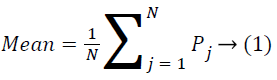
In which, pj is the probability density in iteration j in histogram. N is the total of beans. Two samples of the moment’s analysis in the thermographic images of the healthy person and the person with cancerous masse are indicated in the table 1 as it is clear from the table, the moment difference in the person suffering of cancer and the existence of asymmetry in the healthy person is clear.
| Right breast | Left breast | right breast | Left breast | Moments |
|---|---|---|---|---|
| (non-cancerous) | (non-cancerous) | (cancerous) | (cancerous) | |
| 0.009 | 0.0011 | 0.0009 | 0.0011 | Mean |
| 1.6 | 2.4 | 2.2 | 3.06 | Variance (10-6) |
| 3.4 | 3.7 | 2.2 | 3.7 | Skewness (10-6) |
| 1.2 | 1.1 | 0.1 | 2.1 | Kurtosis (10-8) |
Table 1. Statistical results portraying the mean and variance of Skewness and kurtosis on separated areas.
Table 2 shows the accuracy of the thermography results of the patients compared with pathology outputs for both Hypoechoic and Cystic masses (Figure 2). One of the factors discussed in this research is the subject of asymmetry in thermography. If this factor is detected correctly, it will help the screening and diagnosing of the exact location of the cancer or diseases related to breast tissue. The existence of these factors convinced us that there are complications in breast tissue that must be used in other methods for identifying the exact type of complication. One of the factors that have been addressed in this research is the asymmetry in thermography.
| Thermography Diagnosis | ||||||||
|---|---|---|---|---|---|---|---|---|
| Diagnosing the exact location of the disturbed area | Asymmetry in subjects without single breast | Asymmetry | samples | Symptomatic | ||||
| Number | Percent | Number | Percent | Number | Percent | Total | Percent | |
| 24 | 40 | 35 | 58.33 | 34 | 56.66 | 39 | 65 | hypoechoic mass |
| 11 | 18.33 | 13 | 21.66 | 12 | 20 | 14 | 23.33 | Cystic |
| mass | ||||||||
| 1 | 1.66 | - | - | 1 | 1.66 | 1 | 1.66 | Heterogeneous mass |
| 1 | 1.66 | - | - | 1 | 1.66 | 1 | 1.66 | fibroadenoma |
| 1.66 | - | - | 1 | 1.66 | 1 | 1.66 | Intraductal masses | |
| 1 | 0 | - | - | 1 | 1.66 | 1 | 1.66 | fibro Glandular |
| 0 | 0 | - | - | 1 | 1.66 | 1 | 1.66 | spycoleh masses |
| 0 | 0 | - | - | 1 | 0 | 1 | 1.66 | ISO-echo with cystic |
| 0 | 0 | - | - | 1 | 0 | 1 | 1.66 | Calcified mass |
| 38 | 63.31 | 48 | 79.99 | 51 | 84.99 | 60 | 99.99 | Total |
Table 2. The accuracy of the thermography results based on the type of the tumor.

Figure 2. Presents the thermal images of Hypoechoic, Cystic, Heterogeneous and Fibro glandular mass symptoms.The thermal images of: A- Hypoechoic, B-Cystic, C- Heterogeneous and D- Fibro glandular masses.
According to the magnitude of temperature changes all images were divided in 2 classes; normal (ΔT<1), abnormal (ΔT>1).Out of the 60 females screened, ΔT<1 was observed for 9 cases (3 cases had single breast). Out of these 51 subjects had (ΔT>1). The sensitivity of IR imaging by tumor, age, and size is shown in table 3. Due to results, it is clear that with increasing age, cancer detection will be difficult.
| Characteristic | Samples | Asymmetry | Asymmetry in subjects without single breast | Diagnosing the exact location of the disturbed area | samples |
|---|---|---|---|---|---|
| Age | <30 | 8 | 87% | 87% | 75% |
| 30-40 | 14 | 100% | 100% | 64% | |
| 40-50 | 17 | 82% | 88% | 64% | |
| 50+ | 21 | 66% | 82% | 47% |
Table 3. Sensitivity of IR imaging by age.
The analyzed images can be classified based on their Thermo biological scores into five main groups: TH1-Normal image without vascularity (blood vessels), TH2-Normal image with vascularity, TH3-Equivocal (questionable, but not abnormal), TH4-Abnormal TH5-Very abnormal.
Table 4 presents the results of classifying thermal images on 60 patients based on their Thermo biological scores. It is observed that asymmetry and thermal class are directly related to each other.
| TH5 | TH4 | TH3 | TH2 | TH1 | Number of samples | Symptom |
|---|---|---|---|---|---|---|
| 33 | - | 3 | 2 | 1 | 39 | Hypoechoic mass |
| 12 | - | 2 | - | - | 14 | Cystic mass |
| 3 | - | - | - | - | 3 | Heterogeneous, Fibroadenoma,Intraductal mass |
| 2 | - | - | - | - | 2 | Fibroglandular and Spiculated mass |
| - | - | - | - | 2 | 2 | ISO echo with cystic and calcified mass |
Table 4. Classifying the subject samples based on symptom types and their standard Thermo biological scores.
The statistical analysis as well as the comparison of the stage regarding thermal asymmetry has been shown in table 5. The important point is that the selected cases chosen based on the global article, which is the separation of the core and unnatural edges in the breast tissue it should be among those that suffer cancer, consequently the analysis of specificity and NPV are meaningless in this article.
| NPV | Accuracy | Sensitivity | AbnormalT-Score vs.Normal |
|---|---|---|---|
| 61.53% | 91.89% | 85% | T = -0.5 |
Table 5. The statistical result in analyzing thermal asymmetric on breast.
Discussion
Thermography itself does not provide information about the morphological structures of the breast but it can indicate the functional temperature data and the overall condition of breast tissue vessels [26]. The risk of getting breast cancer has tripled over the bygone half century with respect to modify in way of life and other factors [27]. Therefore it is essential to detect breast cancer primary for better administrator ship of this growing menace. For decades, Medical ultrasound (Sonography) and mammography have been used as the screening methods of select [28].
Conclusion
The risk of breast cancer during the past half century has tripled due to the changes in the people lifestyle and some other factor [25]. The significance of this study is due to the fact that there has been no comprehensive review and comparison done in diagnosing the diseases of breast tissue to relate thermography and ultrasound. Findings from the study indicate that thermography has either advantages or disadvantages in the detection of breast disease. With the advent of a new generation of infrared detectors, infrared thermal imaging has become a medical diagnostic tool for measuring the thermal pattern of abnormal areas. Furthermore, features of Thermal imaging include Sensitivity to the temperature, spatial resolution, and no contact, safe. Thermal images can be stored digitally and then processed by using various software packages and the researcher obtained a comprehensive understanding from the thermal model. The results of this study show that thermography can be used for the early detection and rapid screening. To complete this procedure, methods such as ultrasound can be employed. It can also be considered as a complementary method for ultrasound procedure.The next point is that asymmetry plays a key role in the early detection that we can recognize by means of the basic settings of the camera.Also, Thermography is suitable compared to the ultrasound diagnosis in detecting the breast tissue diseases such as cystic masses and hypo echo masses by adopting asymmetry technique. But this method has some weaknesses in detecting the exact location of the masse, such as cystic. However, thermography is weak in the asymmetry technique and detection of the exact location related to masses as micro calcification. And important point is that, people who once suffered from cancer and had the contents of their infected breast tissue evacuated, the use of asymmetry techniques would be inefficient in thermography. This study emphasizes that thermography should not be used for the first time diagnosis. Considering the progress of this technology and increasing the patient’s requests for a screening method with low price and no ionization, thermography as a breast imaging method can have many potential requests. This technology needs accurate clinical evaluation and it is likely that thermography can be a part of breast screening, detecting and etc. in near future.
References
- Kelly KM, Dean J, Comulada WS, Lee S-J. Breast cancer detection using automated whole breast ultrasound and mammography in radiographically dense breasts. European radiology 2010; 20: 734-742 .
- Saika K, Sobue T. Epidemiology of breast cancer in Japan and the US. JMAJ 2009;52:39-44 .
- Araújo MC, Lima RC, De Souza RM. Interval symbolic feature extraction for thermography breast cancer detection. Expert Systems with Applications 2014;41:6728-6737 .
- Acharya UR, Ng EYK, Tan JH, Sree SV. Thermography based breast cancer detection using texture features and support vector machine. Journal of medical systems 2012;36:1503-1510 .
- Lahiri B, Bagavathiappan S, Jayakumar T, Philip J. Medical applications of infrared thermography: a review. Infrared Physics & Technology 2012;55:221-235 .
- Ng E-K. A review of thermography as promising non-invasive detection modality for breast tumor. International Journal of Thermal Sciences. 2009;48:849-859 .
- Watmough D, Fowler PW, Oliver R. The thermal scanning of a curved isothermal surface: implications for clinical thermography. Physics in medicine and biology 1970;15:1 .
- Steketee J. Spectral emissivity of skin and pericardium. Physics in medicine and biology 1973; 18:686 .
- Collett AE, Guilfoyle C, Gracely EJ, Frazier TG, Barrio AV. Infrared Imaging Does Not Predict the Presence of Malignancy in Patients with Suspicious Radiologic Breast Abnormalities. The breast journal 2014;20:375-380 .
- Nicandro CR, Efrén MM, María Yaneli AA, Enrique MDCM, Héctor Gabriel AM, Nancy PC, Alejandro GH, Guillermo de Jesús HR,RocíoErandi BM. Evaluation of the diagnostic power of thermography in breast cancer using bayesian network classifiers. Computational and mathematical methods in medicine 2013;2013 .
- Vreugdenburg TD, Willis CD, MundyL, Hiller JE. A systematic review of elastography, electrical impedance scanning, and digital infrared thermography for breast cancer screening and diagnosis. Breast cancer research and treatment 2013;137:665-676 .
- Kontos M, Wilson R, Fentiman I. Digital infrared thermal imaging (DITI) of breast lesions: sensitivity and specificity of detection of primary breast cancers. Clinical radiology 2011;66: 536-539 .
- Wishart G, Campisi M, Boswell M, Chapman D, Shackleton V, Iddles S, Hallett A, Britton
- PD. The accuracyof digital infrared imaging for breast cancer detection in women undergoing breast biopsy. European Journal of Surgical Oncology (EJSO) 2010;36:535-540 .
- Arora N, Martins D, Ruggerio D, Tousimis E, Swistel AJ, Osborne MP, Simmons RM. Effectiveness of a noninvasive digital infrared thermal imaging system in the detection of breast cancer. The American Journal of Surgery 2008;196:523-526 .
- Parisky Y, Sardi A, Hamm R, Hughes K, Esserman L, Rust S, CallahanK. Efficacy of computerized infrared imaging analysis to evaluate mammographically suspicious lesions. American Journal of Roentgenology. 2003;180: 263-269 .
- Ng EYK, Ung LN, Ng FC, Sim LSJ.Statistical analysis of healthy and malignant breast thermography. Journal of medical engineering & technology 2001; 25: 253-263.
- Negri S, Bonetti F, Capitanio A, Bonzanini M. Preoperative diagnostic accuracy of fine‐needle aspiration in the management of breast lesions: Comparison of specificity and sensitivity with clinical examination, mammography, echography , and thermography in 249 patients. Diagnostic cytopathology 1994;11:4-8 .
- Goldberg IM, Schick PM, Pilch Y, Shabot MM. Contact plate thermography: a new technique for diagnosis of breast masses. Archives of Surgery 1981;116:271-273 .
- Ciatto S, Palli D, RossellidTM, Catarzi S. Diagnostic and prognostic role of infrared thermography. La Radiologiamedica 1987;74:312-315 .
- Keyserlingk J, Ahlgren P, Yu E, Belliveau N. Infrared Imaging of the Breast: Initial Reappraisal Using High‐Resolution Digital Technology in 100 Successive Cases of Stage I and II Breast Cancer. The Breast Journal 1998;4:245-251 .
- Sterns EE, Curtis AC, Miller S, Hancock JR. Thermography in breast diagnosis. Cancer 1982;50: 323-325 .
- Ohashi Y, Uchida I. Applying dynamic thermography in the diagnosis of breast cancer. Engineering in Medicine and Biology Magazine, IEEE 2000;19:42-51 .
- Zore Z BI, Stanec M, Orešić T, FilipovićZore I. Influence of hormonal status on thermography findings in breast cancer. ActaClinicaCroatica 2013;52:35-42 .
- Sella T, Sklair-Levy M, Cohen M, Rozin M, Shapiro-Feinberg M, Allweis TM, Libson E, Izhaky D. A novel functional infrared imaging system coupled with multiparametric computerised analysis for risk assessment of breast cancer. European radiology. 2013; 23:1191-1198 .
- Muffazzal R, Poonam M, Rajkumar M, Khan F, Sapnak S, Gupta P.K, Jain B. Evaluation of digital infrared thermal imaging as an adjunctive screening method for breast carcinoma: A pilot study. International Journal of Surgery 2014;12:1439-1443 .
- Moghbel M, Mashohor S. A review of computer assisted detection/diagnosis (CAD) in breast thermography for breast cancer detection. Artificial Intelligence Review 2013;39:305-313 .
- Long E, Beales IL. The role of obesity in oesophageal cancer development. Therapeutic advances in gastroenterology 2014;7:247-268 .
- Penhoet EE, Petitti DB, Joy JE. Saving Women's Lives:: Strategies for Improving Breast Cancer Detection and Diagnosis: National Academies Press; 2005 .