ISSN: 0970-938X (Print) | 0976-1683 (Electronic)
Biomedical Research
An International Journal of Medical Sciences
Research Article - Biomedical Research (2016) Volume 27, Issue 4
Enhancement of coronary artery using image fusion based on discrete wavelet transform
Department of Electronics & Instrumentation Engineering, KLN College of Engineering, India
- *Corresponding Author:
- Umarani Durai
Department of Electronics & Instrumentation Engineering
KLN College of Engineering
India
Accepted date: March 30, 2016
Atherosclerosis is called as a systemic disease of the vessel wall that occurs in coronary arteries. The atherosclerotic plaques may cause the narrowing (stenosis) of coronary arteries or complete occlusion of the arteries that leads to heart attack and strokes. X-ray angiography has extremely assisted in the diagnosis of atherosclerosis in patients. During image acquisition the images acquired are degraded due to the presence of noises and artefacts and the clinician find very difficult to diagnosis the stenosis. Therefore there is a need to improve the quality of the image for the easy diagnosis of stenosis. This paper focuses on enhancement of coronary artery blood vessels by mono modal image fusion techniques based on discrete wavelet transform for the easy diagnosis of stenosis. Peak signal to noise ratio (PSNR) computed for a different wavelet filter was used to evaluate the performance of the proposed method. The features are effectively preserved and the simulated result shows that this method enhances the quality of the image in a better way.
Keywords
Image enhancement, Discrete wavelet transform, Image fusion, Histogram equalization.
Introduction
The combination of two images to obtain a single image is known as Medical image fusion. To extract the significant information from the image the image fusion is done. On a single image the current algorithms used for image enhancement work was limited by the role of sensors that is chosen, which fail to offer the essential enhancement's parameters. Subsequently image fusion techniques based on wavelet transform was employed to visualize the blood vessels. The informative features are extorted from the image to improve the clinical diagnosis. The image fusion not only obtains a more accurate and complete description of the image, but also reduces the uncertainty to increase the clinical applicability of image-guided diagnosis and evaluation of medical problems. The main problems in the production of X-ray Angiography images are Low contrast and poor quality. Application of image enhancement technique to the original image is the only solution. A single image approach usually fails in providing the necessary enhancement either due to design or due to observational constraints. By using, the information gathered from multiple images the other possible approach is can be used to enhance image features. One can successfully capture all the relevant information by combining the images. Hence, image enhancement using mono modal temporal image fusion based on 2D discrete wavelet transform is proposed. The DWT method retains the sharp contrast information from all source images and provides both spatial and frequency localization. Soft threshold is used to reduce noise in different sub images obtained after fusion. Even then, the quality of the image is not improved and as a result histogram equalization to the fused image is applied to improve the quality of the image. Peak signal to noise ratio (PSNR) computer for a different wavelet filter was used to evaluate the performance of the proposed method. The features are effectively preserved and the simulated result shows that this method enhances the quality of the image in a better way.
Literature review
In literature and over the past decade different image fusion methods have been offered, a substantial quantity of research has been acquitted on the application of wavelet transform. There are two main image fusion technique, spatial field and frequency-domain. A method based on diverse, focusing to obtain the information from the combination of images in spatial domain was proposed by Li et al. [1]. Gunatilaka et al. and Kor et al. presented an image fusion method based on feature level and pixel level. An image fusion method based on statistical signal processing analysis for weapon detection was presented by Yang and Petrovic et al. proposed multi resolution image fusion based on gradient pixel level image fusion [2-5]. The spatial domain techniques are straightly applied on the source images. There is a numerous of spatial domain approach. The simplest spatial domain approach is the weighted average method. Intensity-hue-saturation (IHS), principal component analysis (PCA), and the Brovey transforms methods are the other methods. Though the spatial domain produces high quality of the image and produce high spectral information they suffer from spectral degradation. Li et al. extended his research by developing an intelligent method of image fusion using artificial neural network (ANN) [1]. To perform ANN, the sample images are required and it does not have an appealing characteristic. Moreover, it is time consuming and it produces more distortions in the images due to which the image gets degraded. As the actual image contains certain structures at different scales or resolutions, a special technique such as multiscale techniques based on the frequency domain can be used for image fusion. Because the medical images are mostly of poor contrast, the spatial information should be kept in the medical images without introducing any distortion or interference. These demands of medical images are better preserved in frequency-domain fusion.
During frequency-domain techniques, images are first decomposed into a series of multiscale coefficients. A variety of fusion rules are used to select the appropriate coefficients and are synthesized through inverse transforms to have the fused image. In recent times, frequency domain techniques have been developed using multiscale transformation, including, Laplacian pyramid transforms, gradient pyramid transform, filter-subtract-decimate pyramid transforms, discrete wavelets transform (DWT), and complex wavelets transform (CWT). Using wavelet transform various researches on image fusion were done, Do et al. proposed contourlet transform, which can give an optimal representation of contours and has been effectively used for image fusion in to eliminate the shortcomings of the wavelet transform [6,7]. Later, Da Cunha et al. describe the complete theory and design of contourlet transform to implement out any application. An image fusion method based on wavelet method for multisensor images was proposed by Pradhan et al. [8,9]. An image fusion method for a multimodal image using wavelet transform was presented by Alfano et al. [10]. A simple DWT-based medical image fusion, which conforms to the weighted fusion rules, has been presented by Cheng et al. [11]. The contourlet transform described by Do et al. possesses shift-invariance thereby suppressing pseudo-Gibbs phenomena [12]. To eliminate this effect an image fusion method based on some modified Laplacian in the frequency domain was presented by Qu et al. [13]. Multimodal image fusion based on wavelet transform was proposed by Yang et al. [14,15]. Dasarathy, Singh et al. and James et al. present a review on medical image fusion [16-19]. Balasubramaniam & Ananthi focused the image fusion using fuzzy sets [20]. Since the fuzzy sets have certain constraints such as are complex in nature, computationally costly and it is limited in exposing the fine structures in the images this method is not suitable for image vessel enhancement. The aforementioned literature survey showed that all the earlier methods were implemented without using any preprocessing techniques. Hence the proposed method uses wavelet domain fusion as an enhancement technique to visualize the coronary artery vessels in X-ray angiography images.
Materials and Methods
The images are acquired through on a Toshiba C arm machine which is outfitted with an image intensifier and a digital flat panel detector the imaging modality provides single plane (2D) fluoroscopic cine window. At 15 to 30 frames per sec (fps) the scans are performed. In the Digital Imaging and communication in medicine (DICOM) format the data sets were saved. It is a medical standard used in most modalities for transfer of images, videos and other diagnostic information. Multiple projections are obtained during angiography of native vessels and graft, including Left anterior oblique (LAO), Right anterior oblique (RAO), frontal, lateral and cranial and caudal views, for optimal visualization. With a C-Arm Machine twenty data sets were acquired. For both scanners a tube voltage of 85 kV and a tube current of 450 to 500 mA were used. All data sets were acquired with ECG-pulsing. A series of 512 × 512 16 bit gray scale images with 256 different gray levels (0-255) are produced as a result of image acquisition.
Results and Discussion
The proposed algorithm is applied to X-ray angiography frames for vessel enhancement through image fusion technique. Since clinical coronary angiogram images are of poor contrast, more detailed and relevant information should be preserved. On a real data set collected from the Government Rajaji Hospital, cardiology section, Madurai, India the performance of the proposed method was tested for five different patient images such as image 1, image 2, image 3, image 4, image 5 and image 6. To preserve the features in the images and to improve the quality of the image the proposed method is carried out along with the histogram equalization. The denoised and artefacts eliminated images are used as the input images. The wavelet filter used for decomposition is biorthogonal (bior2_2), biorthogonal (bior4_4) and Haar wavelet filter.
Figure 1a and Figure 1b shows the fused image using the two wavelet filter. The fused images are obtained with higher-frequency decomposition, and hence the sub images are affected by noises during the fusion of the image. To eliminate the noises soft threshold is applied. Subsequently histogram equalization was applied to the fused image to improve the quality of the image. The Figure 2 depicts fused image equalization using Haar wavelet for the input images. Figures 3a and 3b shows the fused histogram equalized image using Bior and Haar wavelet.

Figure 1: Output of image fusion (a) Bior wavelet transform (b) Haar wavelet.

Figure 2: Output of fused image equalization (a)-(f).

Figure 3: Output of equalized image and its Histogram (a) Bior wavelet (b) Harr wavelet.
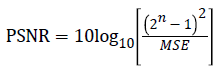
The performance of the proposed system is evaluated by estimating quality metric parameter such as Mean, standard deviation, Peak signal to noise ratio (PSNR) and Entropy. Peak signal to noise ratio (PSNR) is used to measure the quality of a reconstructed image. It is defined as the ratio between the maximum value of an image and the magnitude of background noise. The similarity between the two images was indicated by PSNR. Higher value of PSNR denotes the high quality of the image and it is measured in decible (dB). PSNR is represented by the equation
 → (1)
→ (1)
where MSE=Mean square error
Table 1 shows the comparative performance of histogram equalization on a fusion image at a decomposition level of 4 with Conventional histogram equalization. From the table, it is found that the PSNR of the proposed method using Haar wavelet is high compared to the other wavelet and therefore, the quality of the image is said to be increased.
| Image | Original image | Histogram Equalization | Fusion based histogram equalization | ||||
|---|---|---|---|---|---|---|---|
| PSNR (dB) | Entropy | PSNR(dB) | Entropy | bior 2_2 wavelet | Harr wavelet | Entropy | |
| PSNR (dB) | PSNR (dB) | ||||||
| Image 1 | 24.54 | 5.62 | 26.91 | 5.88 | 43.85 | 45.71 | 5.77 |
| Image 2 | 28.87 | 5.42 | 30.69 | 5.72 | 44.52 | 45.03 | 5.8 |
| Image 3 | 26.89 | 5.61 | 29.78 | 5.75 | 44 .86 | 44.97 | 5.87 |
| Image 4 | 27.97 | 5.61 | 29.81 | 5.65 | 45.35 | 45.79 | 5.67 |
| Image 5 | 24.44 | 5.61 | 28.78 | 5.65 | 43.8 | 44.07 | 5.72 |
| Image 6 | 28.67 | 5.61 | 28.15 | 5.62 | 43.79 | 44.56 | 5.73 |
Table 1: Comparative performance of histogram equalization with conventional histogram equalization and Fusion based equalization for a decomposition level of 4.
The quality estimation Metrics are used to compute the performance of Image fusion using different DWT is shown in the Table 2. Each has its own importance in evaluating the image quality. The entropy and the PSNR of the synthesized image showed an increase in value when the image is decomposed with various levels.
| Image Source | Wavelet Type | Mean | Standard Deviation | Entropy | PSNR(dB) |
|---|---|---|---|---|---|
| Image 1 | Bior 2_2 | 115.19 | 20.48 | 5.04 | 43.85 |
| Bior 4_4 | 115.05 | 20.32 | 5.50 | 43.78 | |
| Haar | 115.19 | 20.48 | 5.77 | 45.71 | |
| Image 2 | Bior 2_2 | 68.38 | 14.38 | 5.24 | 44.52 |
| Bior 4_4 | 68.25 | 14.32 | 5.68 | 44.48 | |
| Haar | 68.54 | 14.38 | 5.80 | 45.03 | |
| Image 3 | Bior 2_2 | 150.01 | 25.26 | 5.19 | 44.86 |
| Bior 4_4 | 149.99 | 25.14 | 5.71 | 44.72 | |
| Haar | 150.09 | 25.26 | 5.87 | 44.97 | |
| Image 4 | Bior 2_2 | 68.91 | 20.36 | 5.26 | 45.35 |
| Bior 4_4 | 68.87 | 20.36 | 5.43 | 45.29 | |
| Haar | 68.90 | 20.24 | 5.67 | 45.79 | |
| Image 5 | Bior 2_2 | 125.60 | 33.34 | 5.32 | 43.80 |
| Bior 4_4 | 125.58 | 33.32 | 5.66 | 43.61 | |
| Haar | 125.60 | 33.34 | 5.73 | 44.07 | |
| Image 6 | Bior 2_2 | 142.60 | 27.36 | 5.15 | 43.79 |
| Bior 4_4 | 142.57 | 27.28 | 5.69 | 43.54 | |
| Haar | 142.60 | 27.36 | 5.73 | 44.56 |
Table 2: Quality evaluation metrics to evaluate the performance of image fusion using DWT.
Conclusion
The investigational consequences showed that the fusion algorithm by the proposed method gives encouraging results. Since the denoised images were used as the input images for image fusion the process did not introduce any distortion to the original image. From the simulation outcomes it is obvious that the consequent fused image and contrast enhanced image consists of information free from unwanted artefacts or distortion, which helps in clinical diagnosis. The statistical analysis obtained using Haar wavelet decomposition has comparatively better performance than the information obtained using the other wavelets. The entropy of the image decomposed at higher levels has greater value. Both these parameters suggest that the decomposed image has more diagnostic information content than the input images.
References
- Li S, Kwok JT, Wang Y. Multifocus image fusion using artificial neural networks. Pattern Recognition Letters 2002; 23: 985-997.
- Gunatilaka AH, Baertlein BA. Feature-level and decision-level fusion of non-coincidently sampled sensors for land mine detection. IEEE Transactions on Pattern Analysis and Machine Intelligence 2001; 23: 577-589.
- Kor S, Tiwary U. Feature level fusion of multimodal medical images in lifting wavelet transform domain. IEEE International Conference of the Engineering in Medicine and Biology Security, 2004; 1479-1482.
- Yang J, Blum RS. A statistical signal processing approach to image fusion for conceled weapon detection’, in Proceedings of the International Conference on Image Processing 2002; 1: 513-516.
- Petrovic VS, Xydeas CS. Gradient-based multiresolution image fusion. IEEE Transactions on Image Processing 2004; 13: 228-237.
- Li S, Kwok JT, Wang Y. Multifocus image fusion using artificial neural networks. Pattern Recognition Letters 2002; 23: 985-997.
- Do MN, Vetterli M. The contourlet transform: an efficient directional multiresolution image representation. IEEE Transactions on Image Processing 2005; 14: 2091-2106.
- Da Cunha AL, Zhou J, Do MN. The nonsubsampled contourlet transform theory, design, and applications. IEEE Transactions on Image Processing 2006; 15: 3089-3101.
- Pradhan PS, King RL, Younan NH, Holcomb DW. Estimation of the number of decomposition levels for a wavelet-based multiresolution multisensor image fusion. IEEE Transactions on Geoscience and Remote Sensing 2006; 44: 3674-3686.
- Alfano B, Ciampi M, Pietro GD. A wavelet-based algorithm for multimodal medical image fusion in Semantic Multimedia. Springer 2007; 117-120.
- Cheng S, He J, Lv Z. Medical image of PET/CT weighted fusion based on wavelet transform. Proceedings of the 2nd International Conference on Bioinformatics and Biomedical Engineering 2008; 2523-2525.
- Do MN, Vetterli M. The contourlet transform: an efficient directional multiresolution image representation. IEEE Transactions on Image Processing 2005; 14: 2091-2106.
- Qu XB, Yan JW, Yang GD. Multifocus image fusion method of sharp frequency localized Contourlet transform domain based on sum-modified-Laplacian. Optics and Precision Engineering 2009; 17: 1203-1212.
- Yang Y. Multimodal medical image fusion through a new DWT based technique. Proceedings of the 4th International Conference on Bioinformatics and Biomedical Engineering 2010; 1-4.
- Yang Y, Huang SY, Gao FJ. Multi-focus image fusion using an effective discrete wavelet transform based algorithm. Measurement Science Review 2014; 14: 102-108.
- Yang Y, Park DS, Huang S, Rao N. Medical image fusion via an effective wavelet-based approach’, Eurasip Journal on Advances in Signal Processing 2010; 1-13.
- Dasarathy BV. Editorial Information fusion in the realm of medical applications-a bibliographic glimpse at its growing appeal. Information Fusion 2012; 13: 1-9.
- Singh R, Khare A. Multiscale medical image fusion in wavelet domain. The Scientific World Journal 2013; 1-10.
- James AP, Dasarathy BV. Medical image fusion: a survey of the state of the art Information Fusion 2014; 19: 4-19.
- Balasubramaniam P, Ananthi VP. Image fusion using intuitionistic fuzzy sets. Information Fusion 2014; 20; 21-30.