ISSN: 0970-938X (Print) | 0976-1683 (Electronic)
Biomedical Research
An International Journal of Medical Sciences
- Biomedical Research (2015) Volume 26, Issue 4
Effects of occlusal hypofunction and its recovery on morphogenesis of molar roots in mice
Orthodontic Science, Division of Oral Sciences, Graduate School, Kanagawa Dental University.
- *Corresponding Author:
- Toshitsugu Kawata
Orthodontic Science, Division of Oral SciencesGraduate School, Kanagawa Dental University, Japan.
Accepted date: June 08 2015
The effects of long-term, artificially created, hypofunctional occlusion and its recovery on the morphology of the first molar root in mice have been investigated. C57BL/6J (Jackson Laboratory, Bar Harbor ME, USA) mice were randomly divided according to their periodontal conditions into normal, hypofunctional, and recovery groups (n = 4). In the experimental hypofunctional and recovery groups, a bite-raising appliance was set to produce hypofunction at the molar region. All groups were analyzed at 16 weeks of age using a light microscope. Root length, width, and area as well as the thickness and the area of the periodontal ligament (PDL) space of the maxillary first molar were calculated. Roots were longer and narrower in the hypofunctional group than in the control group. The mesial root in particular was markedly affected. Root area was significantly smaller in the hypofunctional group than in the other groups. PDL thickness and area were also significantly less in the hypofunctional group than in the control group, but were greater in the recovery group than in the hypofunctional group. These results suggest that root size and the PDL structure may be reduced by disuse atrophy resulting from a defect in occlusal function, but may be recovered following the gain of occlusal stimuli.
Keywords
Hypofunctional occlusion, Occlusal recovery condition, Root morphology, PDL space.
Introduction
Occlusal or masticatory mechanical loading is an important regulatory factor for periodontal tissue homeostasis. The stress of functional occlusion has been suggested to be important for the development and thickening of periodontal collagen fibers [1]. The progressive atrophy of Sharpey's fibers, loss of their regular arrangement, narrowing of the periodontal space, and the of alveolar bone following the lack of occlusal function have been reported previously [2-5].
Histologically, teeth exhibit a hypofunctional periodontium in the absence of occlusal function [6,7]. Periodontal fibers were shown to be thinner, less in number and density, and disordered when the function of the teeth was diminished in mice [3], rats [8]. macaque monkeys [9,10], and humans [6]. Atrophic changes in the periodontal ligament (PDL), such as narrowing of the periodontal space, disorientation of collagen fibers [11], and vascular constriction have been associated with the loss of occlusal function [3,4].
Periodontal hypofunction resulting from the lack of contact with an opposing tooth is encountered in certain malocclusions such as an open bite and ectopic teeth. A longterm hypofunctional condition may be one possible etiological factor for root resorption during orthodontic treatments. The extent of root resorption was shown to be significantly greater for hypofunctional teeth than for control teeth under normal occlusal conditions during experimental tooth movement in rats [12]. Clinical studies also demonstrated that the severity of root resorption, which was examined radiographically, was greater in open bite cases than in deep bite cases before and after orthodontic treatments [13] In histological studies of root morphology, the number of histological sections is typically limited to a small number per tissue sample, and it is impossible to obtain histological sections from different aspects using a single tissue sample. Furthermore, the thickness of the PDL can be easily influenced by tissue preparation, such as handling, decalcification, and dehydration of the specimen [14].
The aim of the present study was to three-dimensionally examine the morphology of mouse molar roots and the PDL space under a long-term hypofunctional nonoccluding condition and compare these results with those obtained under normal occlusion and occlusal recovery conditions. We also attempted to elucidate the relationship between occlusal function and periodontal tissue homeostasis.
Materials and Methods
Experimental Animals
C57BL/6J (Jackson Laboratory, Bar Harbor ME, USA) mice were used in the present study. Water was provided ab libitum and mice were kept in a 12-hour light/dark environment at a constant temperature of 23°C. All animals were fed a powder diet (Rodent Diet CA-1; Japan CLEA Inc, Tokyo, Japan). During the experimental period, the mice were weighed once a week.
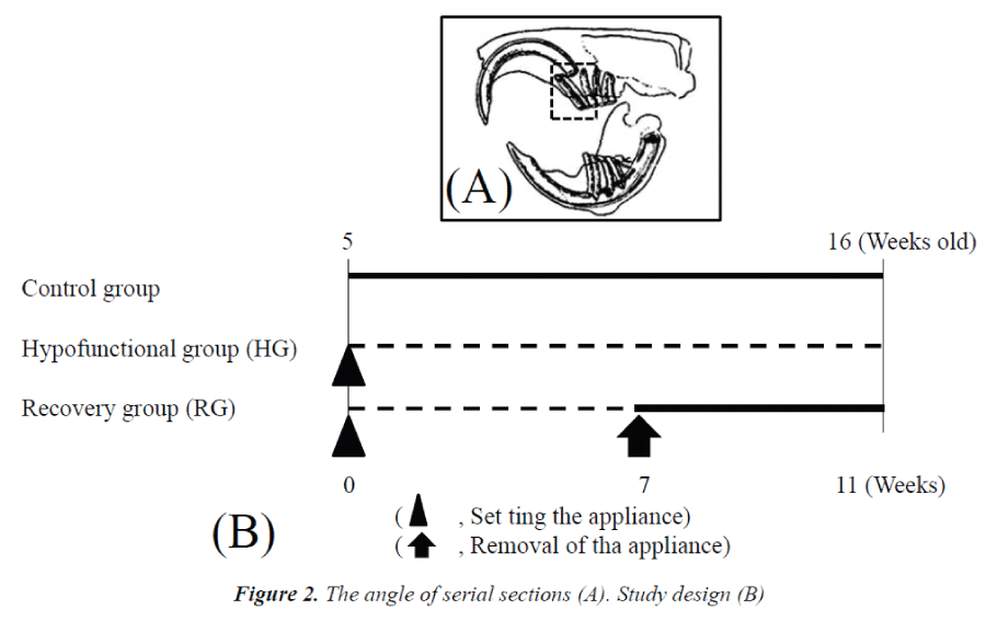
Mice were randomly divided into three groups of four mice each: one untreated control group and two experimental groups. In the experimental hypofunctional and recovery groups, a bite-raising appliance was set to produce hypofunction at the molar region [15]. The appliance consisted of a metal cap made of band material (Tomy International Inc, Fukushima, Japan) and bonded with composite resin (Clearfil Majesty LV; Kuraray Co. Ltd,. Okayama, Japan) on maxillary and mandibular incisors, respectively (Fig. 1). In the recovery group, the appliances were removed 7 weeks after initiation of the experiment in order to recover occlusal contact. Mice were sacrificed at 16 weeks of age. The experimental study design is summarized in Figure. 2.
Morphological Analysis
Root length, width, and area as well as the thickness and area of the PDL space of the maxillary molar were calculated on images using landmarks identified on the roots of the first molar as described below:
Sagittal view.
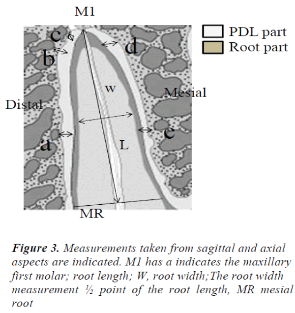
• Root length: measured along the long axis of the root from the cemento-enamel junction (CEJ) and root furcation to the apex (Fig.3).
• Root width: measured perpendicular to the long axis of Long-term study of teeth without antagonist in mice
• mesial (M) roots at the midpoint of each root (Fig. 3).
• Root area: calculated from the CEJ and root furcation to the apex (Fig. 3).
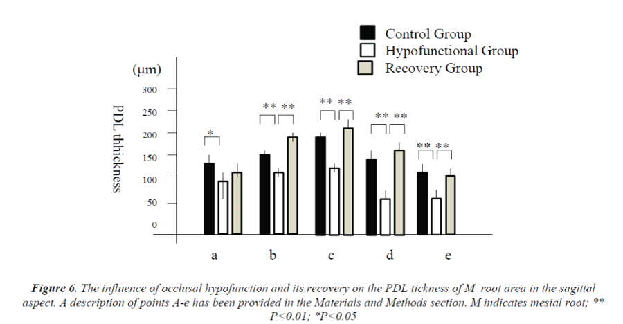
• PDL thickness: recorded from the distance from the root surface to the alveolar bone of M roots at five points. Two perpendicular lines to the long axis of the root divided the root into middle (a, e) and apical (b, d) thirds. Point c was located at the root apex (Fig. 3).
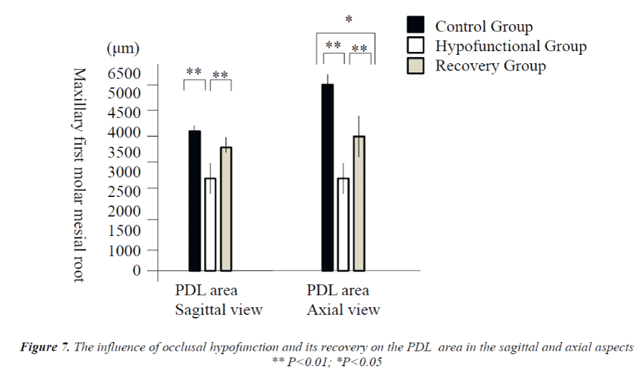
• PDL area: The radiolucent area between the root and the alveolar bone was calculated in sagittal and axial slices (Fig. 3).
Axial view
• Root area in the axial aspect: The root area on a cross-sectional plane passing through the middle point of each root: M, mid-buccal (Fig. 3).
The same investigator performed all measurements, and every measurement was repeated three times. The mean value was used as the final measurement. To assess measurement reproducibility, measurements were performed 10 times for one randomly selected root. The standard error was limited to less than 1%.
Statistical Analysis
Data are expressed as the mean ± standard error of the mean. To determine the significance of differences among groups of mice, we performed a repeated one-way analysis of variance and the Tukey-Kramer test using Statview (Abacus Concepts Inc, Berkeley, Calif). Confidence level P < 0.05.
Results
The Influence of Occlusal Hypofunction and Its Recovery on Root Length and Width of M and DB Roots in the Sagittal Aspect
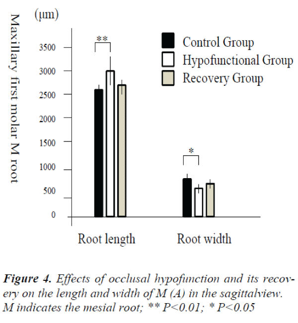
The root in the sagittal aspect was significantly longer (P < 0.01) and narrower (P < 0.05) in the hypofunctional group than in the control group (Fig. 4).
These results showed that roots were slightly longer in the hypofunctional and recovery groups and narrower in the hypofunctional group than in the other groups.
The Influence of Occlusal Hypofunction and Its Recovery on Root Area in the Sagittal and Axial Aspects
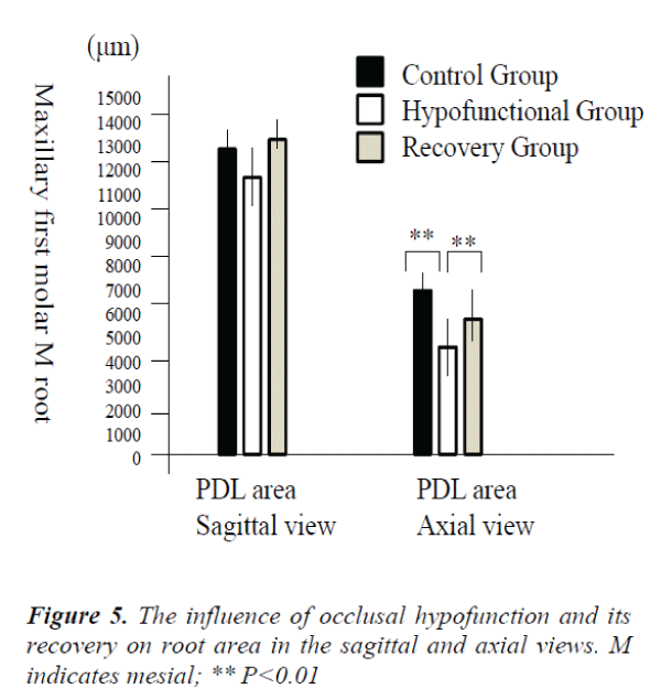
No significant differences were observed in root area between M roots in the sagittal aspect, although the root area of the M root was significantly smaller in the hypofunctional group than in the other groups (P < 0.01; Fig. 5A). The area of the M root in the axial aspect was significantly smaller in the hypofunctional group than in the other groups (Fig.5). These results show that the M root was significantly smaller in the hypofunctional group than in the other groups.
The Influence of Occlusal Hypofunction and Its Recovery on the PDL Thickness of M and DB Roots in the Sagittal Aspect
The PDL space of the M root was significantly narrower in the hypofunctional group than in the control group at points a, c, d, and e, and in the recovery group at points b, c, d, and e (Fig.6). Moreover, the PDL was narrower in the recovery group than in the control group at point a (P < 0.01; Fig.6).
These results demonstrated that PDL thickness was slightly less in the hypofunctional group than in the control group, but increases in the recovery group compared to the hypofunctional group.
The Influence of Occlusal Hypofunction and Its Recovery on the PDL Area in Sagittal and Axial Aspects
The PDL areas around the M roots in the sagittal aspect were significantly smaller in the hypofunctional group than in the control group, and the PDL area of the M root was significantly smaller in the hypofunctional group than in the recovery group (P < 0.01; Fig. 7). In the axial view, the area of the PDL around of the maxillary molar M was significantly smaller in the hypofunctional group than in the control group, and the PDL areas around the M roots were significantly smaller in the hypofunctional group than in the recovery group. Moreover, the PDL areas around the M roots were significantly smaller in the recovery group than in the control group (Fig. 7).
These results showed that the PDL area was smaller in the hypofunctional group than in the control group, but became larger in the recovery group than in the hypofunctional group following the reestablishment of occlusal function.
Micro-CT Images of Upper Molar Teeth in Normal Occlusion, Hypofunctional, and Recovery Groups
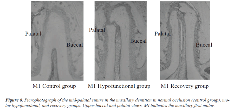
The experimental molars in the hypofunctional group showed greater eruption above the occlusal plane than those in the control and recovery groups. Occlusal hypofunction resulted in less alveolar bone than that in the control group, which appeared to return to normal levels in the recovery group (Fig. 8).
Discussion
The present study was designed to investigate the influence of occlusal or masticatory mechanical stimuli on the morphology of mous molars and the PDL structure. We established an experimental hypofunctional model in the molar region using a bite-raising appliance [15]. This method allowed us to simulate a hypofunctional condition in the molar region and re-establish normal occlusion following removal of the appliance. Clinically, a tooth without its antagonists will elongate towards the opposing crown; this elongation has also been demonstrated experimentally [8,16,17]. In the present study, micro-CT images of the normal and hypofunctional groups and of groups recovering from hypofunctional occlusion suggested that the loss of occlusal contact followed by extrusion f the tooth were associated with root elongation of different degrees in the hypofunctional group; however, this decreased significantly in the recovery group. The results of this study indicated that roots are both shortened by apical root resorption and intruded by functional loads during the recovery period. On the other hand, occlusal hypofunction resulted in a less alveolar bone than that in the control group; however, this level appeared to return to normal in the recovery group. In this regard, occlusal function ensures adequate tooth support. Although bone resorption in the alveolar bone crest occurred during the hypofunctional period when this homeostasis was disrupted; alveolar bone was restored almost to the level of control groups during the recovery period to adapt to the new functional condition. In addition, the loss of attrition was only observed in the hypofunctional group, which suggested that the maxillary molars had no occlusal contact throughout the experimental period.
Atrophic changes in the PDL have been associated with the loss of occlusal function [11,18]. Such changes include narrowing of the periodontal space, disorientation of collagen fibers [3], vascular constriction, and deformation of the mechanoreceptor structure [4]. Other studies using occlusal recovery models reported the widening of blood vessels in the PDL following the application of occlusal stimuli [8,19]. However, it is difficult to adapt these occlusal recovery models, which were simulated in 7-week-old rats, to a clinical situation, because the eruption of the first molars in these rat models was almost finished at 5 weeks old; these rats could naturally bite between 5 and 7 weeks of age before short-term hypofunctional condition was simulated. Previous studies in male rats showed that alveolar bone turnover and osteoblastic/ osteoclastic activity decreased with age [20-22]. In our study, the durations of the hypofunctional condition were 11 and 7 weeks for the hypofunctional and recovery groups, respectively. However, since the growth potential of 12-week-old mice is minimal, it was considered that there was no difference was considered to exist between 7-week and 11-week hypofunctional conditions.
Regarding the influence of occlusal hypofunction and its recovery on the length and width of M roots evaluated in the sagittal aspect, we demonstrated that the M root was significantly longer and narrower in the hypofunctional group than in the control group. Moreover, no significant differences were observed in root areas, except for the M root, which was significantly smaller in the hypofunctional group than in other groups. In this study, we found that the mesial root was mainly affected by hypofunction. This may have been due to the larger size of the mesial root than the rest of the roots. Therefore, changes in the mesial root were clear and easy to quantify.
These results, suggest that the roots of mouse molars with the loss of occlusal stimuli elongate toward the opposing crown, and, since the midpoint of the elongated root in the hypofunctional group shifted towards the apex, root width and area decreased.
In the present study, we demonstrated from most of the PDL measurements that the PDL was significantly narrower in the hypofunctional group than in the control group and that it was wider in the recovery group than in the hypofunctional group [23-25]. The same results were observed for the PDL area. These results strongly suggest that atrophic changes in PDL structure were induced by the loss of occlusal function, but that PDL mechanoreceptors way be recovered, possibly in association with the widening of blood vessels, in the PDL following the application of occlusal stimuli. Moreover, these results suggest that root elongation under the hypofunctional conditions occurred concomitantly with decreases in PDL thickness and tooth eruption.
Conclusion
• Roots were longer and narrower in the hypofunctional group than in the control group. Root area was also significantly smallest in the hypofunctional group than in the other groups.
• PDL thickness and area were significantly less in the hypofunctional group than in the control group, but were greatest in the recovery group than in the hypofunctional group.
• These results suggest that root size and the PDL structure may be reduced by disuse atrophy resulting from a defect in occlusal function, but may be recovered following the gain of occlusal stimuli.
References
- Bernick S. The organization of the periodontal membrane fibres of the developing molars of rats.Arch Oral Biol 1960; 2: 57-63.
- Levy GG, Mailland ML. Histologic study of the effects of occlusalhypofunction following antagonist tooth extraction in the rat. J Periodontol 1980; 51: 393-399.
- Cohn SA. Disuse atrophy of the periodontium in mice.Arch Oral Biol 1965; 10: 909-919.
- Cohn SA. Disuse atrophy of the periodontium in mice following partial loss of function.Arch Oral Biol 1966; 11: 95-105.
- Shimizu Y, Hosomichi J, Kaneko S, Shibutani N, Ono T. Effect of sympathetic nervous activity on alveolar bone loss induced byocclusalhypofunction in rats. Arch Oral Biol 2011; 56: 1404-1411.
- Kronfeld R. Histologic study of the influence of function on the human periodontal membrane. J Am Dent Assoc 1931; 18: 1242-1274.
- Newman WG. Possible etiologic factors in external root resorption. Am J Orthod 1975; 67: 522-539.
- Saeki M. Experimental disuse atrophy and its repairing process in the periodontal membrane. J StomatolSocJpn 1959; 26: 317-347.
- Anneroth G, Ericsson SG. An experimental histological study of monkey teeth without antagonist.Odontol Rev 1967; 18: 345-359.
- Pihlstrom BL, Ramfjord SP. Periodontal effect of nonfunctionin monkeys. J Periodontol 1971; 42: 748-756.
- Kaneko S, Ohashi K, Soma K, Yanagishita M. Occlusalhypofunction causes changes of proteoglycan content in the rat periodontal ligament. J Periodontal Res 2001; 36: 9-17.
- Sringkarnboriboon S, Matsumoto Y, Soma K. Root resorption related tohypofunctionalperiodontium in experimental tooth movement. J Dent Res 2003; 82: 486-490.
- Harris EF, Butler ML. Patterns of incisor root resorptionbefore and after orthodontic correction in cases with anterior open bites.Am J OrthodDentofacialOrthop1992; 101: 112-119.
- Nakamura Y, Noda K, Shimoda S, Oikawa T, Arai C, Nomura Y, Kawasaki K. Time-lapse observation of rat periodontal ligament during function and tooth movement, using microcomputed tomography. Eur J Orthod2008: 30: 320-326.
- Suhr E, Warita H, Iida J, Soma K. The effect of occlusalhypofunction and its recovery on the periodontal tissues of the rat molar: ED1 immunohistochemical study. Orthodontic Waves 2002; 61: 165-172.
- Kinoshita Y, Tonooka K, Chiba M. The effect of hypofunctionon the mechanical properties of the periodontiumin the rat mandibular first molar.Arch Oral Biol1982; 27: 881-885.
- Fujita T, Montet X, Tanne K, Kiliaridis S. Suprapositionof unopposed molars in young and adult rats. Arch Oral Biol. 2009; 54: 40-44.
- Muramoto T, Takano Y, Soma K. Time-related changes in periodontal mechanoreceptors in rat molars after the loss of occlusal stimuli. Arch HistolCytol2000; 63: 369-380.
- Misawa Y, Kageyama T, Moriyama K, Kurihara S, Yagasaki H, DeguchiT, Ozawa H, Sahara N. Effect of age on alveolar bone turnover adjacent to maxillary molar roots in male rats: a histomorphometric study. Arch Oral Biol 2007; 52: 44-50.
- Koike K. The effects of loss and restoration of occlusalfunction on the periodontal tissues of the rat molar teeth—histopathological and histometrical investigation.J JpnSocPeriodontol 1996; 38: 1-19.
- Nishimoto SK, Chang CH, Gendler E, Stryker WF, Nimni ME. The effect of aging on bone formation in rats: biochemical and histological evidence for decreased bone formation capacity. Calcif Tissue Int1985; 37: 617-624.
- King GJ, Latta L, Rutenberg J, Ossi A, Keeling SD. Alveolar bone turnover in male rats: site- and agespecificchanges. Anat Rec 1995; 242: 321-328.
- Sugiyama H, Lee K, Imoto S, Sasaki A, Kawata T, Yamaguchi K, Tanne K. Influences of vertical occlusaldiscrepancies on condylar responses and craniofacial growth in growing rats. Angle Orthod 1999; 69: 356- 364.

