ISSN: 0970-938X (Print) | 0976-1683 (Electronic)
Biomedical Research
An International Journal of Medical Sciences
- Biomedical Research (2007) Volume 18, Issue 3
Effect of withaferin, a radiosensitizer, on the erythrocyte antioxid-ants in carcinoma of uterine cervix
Reshma K1, Rao A.V.2, Dinesh M3, Vasudevan DM4
1Department of Biochemistry, Kasturba Medical College, Mangalore, Karnataka, India
2Department of Biochemistry, KS Hegde Medical Academy, Mangalore, Karnataka, India
3Department of Oncology, Amrita Institute of Medical Sciences, Cochin, India
4Department of Biochemistry, Amrita Institute of Medical Sciences, Cochin, India
- Corresponding Author:
- Reshma Kumarchandra
Department of Biochemistry Kasturba
Medical College Mangalore, Karnataka
State India
Accepted date: July 31 2007
Withaferin, an active component obtained from the root extracts of Withania somnifera (Ashwaganda), showed antitumor and radiosensitising effects in animals. A similar approach in human cancer patients could probably increase the therapeutic outcome. Antioxidants are good markers of free radical induced tissue damage or in other words, radiosensitisation of tumor cells. Therefore an assay of erythrocyte antioxidant levels namely GSH, GSH-PX,SOD and G6PD were performed in cancers of uterine cervix, before, in the midst of, and post radiation and compared with the erythrocyte levels of the same in normal controls. Although a high level of GSH was observed in the baseline samples, this level was consistent after treatment. A significant decrease in GSHPX values following radiation was observed. SOD and G6PD levels remained non significant. Therefore establishing the role of withaferin as a radiosensitiser seems to be ambiguous.
Keywords
Radiotherapy, Radiosensitiser, Withaferin, Antioxidants
Abstract
Withaferin, an active component obtained from the root extracts of Withania somnifera (Ashwaganda), showed antitumor and radiosensitising effects in animals. A similar approach in human cancer patients could probably increase the therapeutic outcome. Antioxidants are good markers of free radical induced tissue damage or in other words, radiosensitisation of tumor cells. Therefore an assay of erythrocyte antioxidant levels namely GSH, GSH-PX,SOD and G6PD were performed in cancers of uterine cervix, before, in the midst of, and post radiation and compared with the erythrocyte levels of the same in normal controls. Although a high level of GSH was observed in the baseline samples, this level was consistent after treatment. A significant decrease in GSHPX values following radiation was observed. SOD and G6PD levels remained non significant. Therefore establishing the role of withaferin as a radiosensitiser seems to be ambiguous.
Introduction
Cancer of uterine cervix is one of the leading causes of cancer death among women worldwide[1]. Early stages of this disease can be treated by surgery or with radiation. Failure of radiotherapy in the local control of solid tumors is often attributed to the presence of radioresistant hypoxic cells. Therapeutic outcome can be improved by using the chemicals(radiosensitizers) that increase the radiosensitivity of tumor cells, so that a higher tumor killing is achieved at conventional doses of RT. Furans, thiophenes, imidazoles, pyrazoles, pyrroles and thiazoles have been synthesised and tested as hypoxic cell radiosensitizers[2].Withaferin A, the active component obtained from the alcoholic extracts of the dried roots of the plant Withania somnifera showed significant antitumor and radiosensitising effects in experimental tumors induced in mice without any noticeable systemic toxicity [3,4].As radiation kills tumor cells by generating free radicals and since withaferin sensitizes the tumor tissue to radiation we sought to study the influence of the latter on the erythrocyte antioxidant levels.
Materials and Methods
20 cases of carcinoma of uterine cervix (stage 111 B ) were considered for the study. All patients were treated with radiation at Kasturba Medical college, Hospital, Mangalore, India.
Inclusion criteria
All patients selected were aged between 30 and 70 years. All cancer patients were selected based on the karnofsky’s performance scale KPS>70% (10).[Cares for self but unable to carry out normal activity: shows some signs or symptoms of the disease]
The patients had no previous history of treatment and received radiotherapy at a dose of 60 Gy in 30 fractions over 6 weeks.
Exclusion criteria
All patients were subjected to thorough clinical examination and those with severe systemic illness like diabetes mellitus, coronary artery disease and tuberculosis were excluded.
All patients with carcinoma of cervix were treated with withaferin prior to radiotherapy,at a dose of 400mg/m2, 2 hrs prior to each sitting. The Institutional Ethical Committee had approved the drug trials..
Age and sex matched healthy non hospitalized controls (n=25) were considered for the comparative study with the patients.
NADPH, Riboflavin, Lmethionine and Glutathione standard were obtained from SRL company limited.Glucose 6 phoshate was purchased from Loba chem.. Cyanomethemoglobin standard was bought from Ranbaxy.DTNB was obtained from SISCO, NBT from S.D. Fine chem. Ltd. Cumene hydroperoxide from Fluka, Ag L Buchio, Switzerland and Glutathione reductase (E.C.1.6.4.2.) Type111 from Bakers yeast from Sigma chemicals,U.S.A.
Heparinised vacuotainers were purchased from Babul Biomedicals Pvt. Ltd, Ahmedabad.
5 ml of venous blood was collected from patients in three stages:
a. 0 days of radiation (Baseline sample)
b. 15 days of radiation (1 follow up sample)
c. 30 days of radiation (11 follow up sample all patients were not available)
Likewise 5 ml of blood was collected from controls.
Blood samples were kept in an upright position at room temperature for one hour. Once the plasma separated erythrocytes were collected and the hemolysate was prepared by the reported protocol.
This hemolysate was used for estimation of different parameters. Glutathione (GSH) was assayed by the method of Beutlar et al [5]. Glutathione peroxidase (GSH-PX) by the method of Paglia and Valentine [6,7]. Glucose 6 phosphate dehydrogenase (G6PD) was estimated by monitoring the increase in absorbance at 340 nm [8]. Superoxide dismutase (SOD) was assayed by the method of Beauchamp and Fridovich [9,10]. Haemoglobin (Hb) concentration in the RBC was determined by cyanomethemoglobin method [11]. The activities of GSH was expressed as nmol/g Hb and the activities of other antioxidants were expressed in terms of units/g Hb.
Statistical analysis
Kruskal Waalis test was used for comparison between independent groups. Wilcoxons rank sign test was used for comparing the follow up cases.
Results
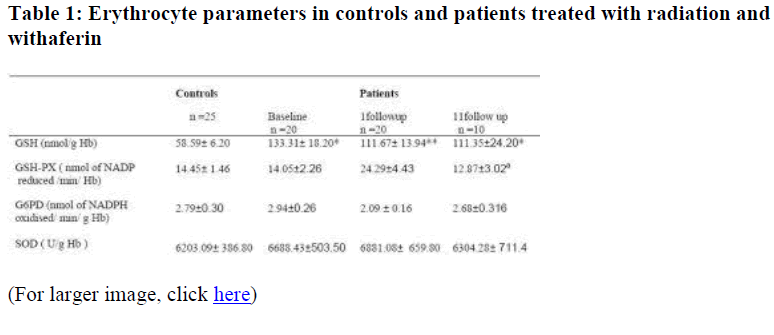
Erythrocyte GSH increased in cancer of uterine cervix as compared to controls p<0.01. Following treatment, a decrease is observed which is not statistically significant but significantly high as compared to controls p<0.05. There were no significant changes when GSH-PX values of patients were compared with controls. However, the values were significantly low in the second follow up as compared to first follow up p<0.05.SOD and G6PD values did not vary between follow up samples or between the two groups.
slortnoc ot derapmoc tnacifingis 10.0>p?
slortnoc ot derapmoc tnacifingis 50.0>p??
a p<0.05 significant between first and second follow up
Discussion
Certain works have reported low levels of GSH in cancer of uterine cervix and cervical intraepithelial neoplasia [12,13]. A fall in GSH concentration in blood plasma in cancer of uterine cervix after one fraction of RT was also observed which correlated positively with tumor response as reported by Jadhav et al [14]. On the contrary, elevated levels of GSH in cancer tissues (oral cancer,lung squamous cell carcinoma,cervical and other squamous cell carcinoma) has been observed by Wong et al [15] which has been attributed to abnormal proliferative activities in cancer tissues. Our work has observed an elevation of GSH in the blood which indicates that the abnormal proliferative activities reflects in the blood as well. However, treatment with radiation and withaferin have not resulted in alteration in the values of GSH.
GSHPX values have decreased in cancer tissues [16] and in the blood of cancer patients [17]. A fall in erythrocyte GSHPX levels was reported in oesophageal cancer [18] advanced gastrointestinal cancer, breast cancer [19], colon cancer [20] and lung cancer [21]. Although we report no change in the baseline samples of cancer patients as compared to controls, RT has led to a fall in the GSHPX levels which could not be reverted by Withaferin. G6PD activities have neither altered in baseline samples nor in the post treated samples as compared to controls which would mean that the reducing equivalents for the generation of reduced glutathione is provided by some other source. SOD levels remained nonsignificant in the pretreated and post treated samples versus controls.
An increase in the antioxidant enzymes namely SOD, GSHPX and catalase was reported in the rat brain following withaferin treatment [22]. No such effects were observed with respect to withaferin in the present work. Therefore, the role of withaferin in sensitizing the cancer tissue to radiation remains obscure.
Acknowledgements
We wish to acknowledge Dr Umadevi, Department of Radiobiology, Kasturba Medical College, Manipal, for having provided with the radiosensitising drug for our study.
We thank the Institutional Ethical Committee for having permitted us to conduct the study.
References
- Shanta V, Krishnamurthy S, Gajalakshmi CK, Swaminathan R, Ravichandran K. Epidemiology of cancer of the cervix:global and national perspective. J Indian Med Assoc 2000; 98: 49-52.
- Fieldon FM, Adams GE, Cole S, Naylor MA, O’Niel P, Stephen MA, Stratford LJ. Assesment of a range of novel nitroaromatic radiosensitizers and bioreductive drugs. Int J Radiat Oncol Biol Phys 1992; 22: 707-711.
- Devi PU, Agakik K, Ostapenko V, Tanaka Y, Sugahara T. Withaferin A. A new radiosensitizer from the Indian medicinal plant Withania somnifera. Int J Radiat Biol 1996; 69: 193-197.
- Devi PU. Withania somnifera Dunal (Ashwagandha) potential plant source of a promising drug for cancer chemotherapy and radiosensitisation. Indian J Exp Biol 1996; 34: 927-932.
- Beutler E, Duron O, Kelley B. Improved method for the determination of blood glutathione. J Lab Clin Med 1963; 61: 882- 888.
- Paglia DE, Valentine WN. Studies on the qualitative and quantitative characterization of glutathione peroxidase. J Lab Clin Med 1967; 70: 158-169.
- Lawrence RA, Burk RF. Glutathione peroxidase activity in seleniumdefecient rat liver. Biochem Biophys Res Commun 1976; 71: 952-958.
- Vorley H, Gowenlock AH, Bell M: In: Practical Clinical Biochemistry, 5th edition,vol 1, CBS Publishers, New Delhi. Pp 730-731.
- Beauchamp C, Fridovich I.Superoxide dismutase: improved assays and an assay applicable to acrylamide gels. Annal Biochem 1971; 44: 276-287.
- Mc Cord JM, Fridovich I. Superoxide dismutase: An enzymatic function of erythrocuprein(hemocuprein). J Biol Chem 1969; 244: 6049-6055.
- Varley H, Gowenlock AH, Bell M. In: Practical Clinical Biochemistry, 5th edition, vol 1 CBS Publishers, New Delhi. Pp 979-980.
- Mukundan H, Bahadur AK, Kumar A, Sardana S. Glutathine level and its relation to radiation therapy in patients with cancer of uterine cervix. Indian J Exp Biol 1999; 37: 859-864.
- Kumar A, Sharma S, Pundir CS, Sharma A. Decreased plasma glutathione in cancer of uterine cervix. Jpn J Clin Oncol 1993; 23: 14-19.
- Jadhav GK, Bhanumathi P, Umadevi P, Seetharamaiah T, Vidyasagar MS, et al. Possible role of glutathione in predicting radiotherapy response of cervix cancer. Int J Radiation Oncology. Biol Phys 1998; 41: 1-3.
- Wong Dy, Hsiao YL, Poon CK, Kwan PC. Glutathione concentration in oral cancer tissues. Cancer. Lett. 1994; 30. 81: 111-116.
- Durak I, Beduk Y, Kavutcu M, Ozturk S. Activities of superoxide dismutase and glutathione peroxidase enzymes in cancerous and noncancerous human kidney tissues. Int Urol Nephrol 1997; 29: 5-11.
- Wasowicz W, Gromadzinska J, Skodowska M, Popadiuk S. Selenium concentration and glutathione peroxidase activity in blood of children with Cancer. J Trace Elem Electrolytes Health Dis 1994; 53-57.
- Li WJ (Selenium content and glutathione peroxidase in erythrocytes from different populations in area with high and low mortality of oesophageal cancer) Chung
- Paw Owicz, Zachara BA, Trafikowska U. Blood selenium concentrations and glutathione peroxidase activities in patients with breast cancer and with advanced gastrointestinal cancer. J Trace Elem Electrolytes Hea-lth Dis 1991; 5: 275-277.
- Jendryczko A, Pardela M, Kozlowski A. Erythrocyte glutathione peroxidase in patients with colon cancer Neoplasm 1993; 40: 107-109.
- Zachara BA, Marchaluk-Wisniewska E, Maciaq A, Peplinski J, Skokowski J, et al. Decreased selenium concentration and glutathione peroxidase activity in blood and increase of these parameters in malignant tissue of lung cancer patients. Lung 1997; 175: 321-332.
- Bhattacharya SK, Satyan KS, Chakraborty A. Effect of Trasina, an ayurvedic herbal formulation on pancreatic slet superoxide dismutase activity in hyperglycemic rats. Indian J Exp Biol 1997; 35: 297-299.
