ISSN: 0970-938X (Print) | 0976-1683 (Electronic)
Biomedical Research
An International Journal of Medical Sciences
Research Article - Biomedical Research (2017) Volume 28, Issue 3
Comparison of the soft tissue orthodontic analysis measurements between conventional lateral cephalograms and CBCT derived lateral cephalograms
Mashallah Khanehmasjedi1*, Amirfarhang Miresmaili2, Sanaz Jafari3 and Samaneh Khanehmasjedi4
1Associate professor, Orthodontics Department, Ahvaz Jundishapur University of Medical Sciences, Ahvaz, Iran
2Associate professor, Orthodontics Department, Hamedan Dentistry Faculty, Hamedan, Iran
3Postgraduate Orthodontic Student, Orthodontics Department, Ahvaz Jundishapur University of Medical Sciences, Ahvaz, Iran
4Dental Student, Ahvaz Jundishapur University of Medical Sciences, Ahvaz, Iran
- *Corresponding Author:
- Mashallah Khanehmasjedi
Associate Professor of Orthodontics
School of Dentistry
Ahvaz Jundishapur University of Medical Sciences,Iran
Accepted date: July 14, 2016
Conventional lateral cephalograms are ordered for patients even with available Cone-Beam Computed Tomography (CBCT) radiographs which may increase the patient’s radiation exposure. According to past studies landmark identification and hard tissue analysis errors on CBCT-derived lateral cephalograms were comparable to those of conventional lateral cephalograms. According to the soft tissue paradigm, diagnosis and treatment planning are based on the soft tissue goals. The aim of this study was to compare the soft tissue analysis between CBCT-derived and conventional lateral cephalograms of the same patient. Twenty-two patients who had both 12-inch CBCT scan (NewTom 3G) and conventional lateral cephalograms available within a 6-month time period were selected. Landmark identification carried out by two operators at the same time for Dolphin imaging software (v11.2). 8 angular and 11 linear soft tissue variables were measured. Paired t-test comparison of two groups revealed no statistically significant differences between the groups except for inclination of nasal base (P<0.05 was considered significant). CBCT-derived lateral cephalograms can be used for the soft tissue analysis as an alternative to conventional lateral cephalograms, when patient’s CBCT volume is already available. This can reduce additional X-ray exposure.
Keywords
Cephalometry, Cone-beam computed tomography, Soft tissue.
Introduction
Cephalometric radiography is one of the most important diagnostic adjuncts in orthodontics. After identifying landmarks, linear and angular measurements are used for the explanation of facial and maxillary and mandibular morphology, deformities, growth prediction, treatment planning, assessment of treatment outcome and research [1]. Computed tomography (CT) is unsuitable for most dental and orthodontic purposes because of high radiation exposure and high expense. Recently cone-beam computed tomography (CBCT) which provides lower radiation exposure than conventional CT is being used increasingly in orthodontics and other fields in dentistry [2-4]. Since conventional lateral cephalograms have provided a 2-dimensional image of a 3- dimensional subject, some superimpositions, magnification and other imaging errors are probable which makes it difficult to compare them with three-dimensional CBCT [5].
It is possible to obtain lateral cephalograms from 3- dimensional CBCT and compare its measurements with those of conventional lateral cephalograms [6-8]. Previous few studies demonstrated some controversies [9,10]. Nalcaci et al., compared two-dimensional and 3-dimensional cephalometric measurements. The results were comparable between two groups except for upper incisor angulation [9]. Bholsithi et al., reported that 3-dimensional measurements are comparable with two-dimensional cephalometric measurements only in midline [10]. Otherwise, Van Vlijmen et al., reported two groups of comparable measurements [4].
Nowadays conventional lateral cephalograms are ordered for patients even with available CBCT radiographs which may increase patient’s radiation exposure. According to the past studies landmark identification and hard tissue analysis errors on CBCT-derived lateral cephalograms are comparable to those of conventional lateral cephalograms [9-11]. According to soft tissue paradigm, new orthodontic approach is based on soft tissue markers in diagnosis and treatment planning [12]. We believed that CBCT-derived lateral cephalograms can be used for soft tissue analysis as an alternative to conventional lateral cephalograms, when patient’s CBCT volume is already available because it reduces the expense and additional X-ray exposure and as there is possibility of skull orientation before generating CBCT-derived lateral cephalograms, it can be considered more reliable than conventional lateral cephalograms. Therefore, the aim of this study was to compare soft tissue analysis between CBCT-derived lateral cephalogram and conventional lateral cephalograms of the same patients.
Materials and Methods
Twenty-two patients who had both CBCT scan (NewTom 3G Verona Italy) with 12 inch (FOV [field of view]) and conventional lateral cephalograms available within a 6-month time period were selected. The study is approved by the ethical committee of the Ahvaz Jundishapur University of Medical Sciences. All patients were aware of the study and signed a written constant. The study was performed during 6 months, from February 2015 to July 2015.
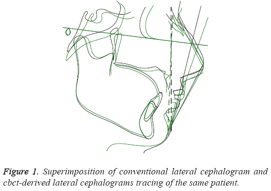
The CBCT scans were imported in Dolphin imaging 3D (The Dolphin 3D software is a powerful tool that makes processing 3D data extremely simple, enabling dental specialists from a wide variety of disciplines to diagnose, plan treatment, document and present cases), Chatsworth Calif version 11.2 by horizontally orientation of the Frankfort and the infraorbital plane. The mid-sagittal plane was oriented vertically. The lateral cephalograms were generated with 9% magnification to simulate the conventional lateral cephalometric radiographs geometry (according to the manufacturer’s instruction). Landmark identification carried out by two operators at the same time (one operator was tracing the landmarks while the other operator observed) for Dolphin imaging software (v11.2) (Figure 1). 8 angular and 11 linear soft tissue variables were measured (Table 1).
| Angular Variables | Linear Variables |
| Nasofacial angle | Maxillary prognathism |
| Inclination of nasal base | Upper lip prominence |
| Nasomental angle | Mandibular prognathism |
| Mentocervical angle | Lower lip prominence |
| Nasolabial angle | Chin prominence |
| Angle of facial convexity | Soft tissue chin thickness |
| Facial angle | Upper lip curvature |
| H-line angle | Upper sulcus depth |
| Lower sulcus depth | |
| Upper lip strain | |
| Upper lip thickness |
Table 1: Angular and linear variables measured in this study.
Statistical analysis
Spss version 20 was used to analyze the data. Paired t-test was used to compare measurement differences in two imaging modalities (conventional lateral cephalograms and CBCTderived lateral cephalograms). P<0.05 was considered significant.
Results
According to paired t-test comparison, two groups revealed no statistically significant differences between the groups except for inclination of nasal base (Table 2).
| Variables | Conventional Lateral Cephalogram | CBCT-derived Lateral Cephalogram | P value | ||
|---|---|---|---|---|---|
| Mean | SD | Mean | SD | ||
| Nasofacial angle | 31.10 | 4.16 | 31.07 | 4.43 | 0.98 |
| Inclination of nasal base | -51.37 | 16.45 | -24.47 | 6.62 | 0.00 |
| Nasomental angle | 129.28 | 7.24 | 128.97 | 7.83 | 0.89 |
| Mentocervical angle | 94.31 | 10.34 | 84.98 | 23.29 | 0.09 |
| Nasolabial angle | 87.18 | 6.71 | 86.12 | 6.48 | 0.59 |
| Angle of facial convexity | 4.11 | 0.28 | 4.88 | 0.68 | 0.77 |
| Facial angle | 90.28 | 4.11 | 90.68 | 4.88 | 0.77 |
| H-line angle | 12.65 | 4.96 | 13.82 | 5.49 | 0.46 |
| Maxillary prognathism | -8.92 | 6.68 | -8.49 | 7.48 | 0.84 |
| Upper lip prominence | -1.83 | 2.64 | -1.40 | 2.17 | 0.54 |
| Mandibular prognathism | 2.82 | 2.64 | 3.35 | 2.49 | 0.49 |
| Lower lip prominence | 3.07 | 0.78 | 2.81 | 0.78 | 1 |
| Chin prominence | -6.65 | 6.97 | -8.82 | 7.78 | 0.33 |
| Soft tissue chin thickness | 13.15 | 2.67 | 14.03 | 3.15 | 0.31 |
| Upper lip curvature | 2.36 | 0.95 | 2.39 | 0.33 | 0.38 |
| Upper sulcus depth | 2.42 | 1.21 | 2.57 | 1.22 | 0.68 |
| Lower sulcus depth | 3.78 | 2.43 | 4.34 | 2.35 | 0.42 |
| Upper lip strain | 13.10 | 2.34 | 12.93 | 4.77 | 0.88 |
| Upper lip thickness | 8.30 | 7.70 | 7.94 | 6.96 | 0.87 |
Table 2: Means and Standard Deviations of Angular and Linear Measurements of two Imaging Modalities and P values of the t-test between them
Discussion
Using conventional lateral cephalograms as they provide twodimensional views of 3-dimensional objects may cause landmark identification problems according to superimposition of other facial structures [13,14]. Other problems such as magnification and other imaging errors may also occur [5]. Using CBCT-derived lateral cephalograms eliminates superimposition and magnification errors in conventional lateral cephalograms. The higher radiation dose of CBCT limits it’s use in dentistry [13]. It seems to be necessary to compare conventional lateral cephalometry analysis norms with CBCT-derived lateral cephalograms. If the results are compatible for patients with available CBCT, there will be no need for further radiation to the patient for obtaining conventional lateral cephalograms.
Past studies demonstrated that landmark identification and hard tissue analysis errors on CBCT-derived lateral cephalograms are comparable to those of conventional lateral cephalograms in the studies of Nalcaci, Bholsithi, Chang, Kumar, Damstra, Zamora, Yitschaky, and Park [7-11,15-18]. According to the present study, cephalometric soft tissue measurements performed on conventional lateral cephalograms are compatible with measurements on CBCT-derived lateral cephalograms except for inclination of nasal base. Bholsithi et al., demonstrated that landmark identification is more comparable for midline landmarks [10]. van Vlijmen et al., revealed that the Measurements on CBCT-constructed cephalometric radiographs are comparable to conventional cephalometric radiographs, and are therefore suitable for longitudinal research [6]. In a study by Farhadian et al., on comparison of cephalometric analysis between conventional and CBCT generated lateral cephalograms it was shown that LC could successfully be replaced by GLC. Because it is possible to select the best orientation of the skull before generating GLC from CBCT DICOM files, GLC could be more reliable than LC [19]. In this study, because of 6-month time period limitation for each patient’s radiographs, growth could not lead to measurement errors.
Differences in the inclination of nasal base between two radiographic approaches could be resulted from following reasons: 1- overall, measuring the inclination of nasal base might be not accurate due to nasal soft tissue mass. 2- Error in defining this parameter in Dolphin software could be considered as a possible factor. Given that this landmark was only the lateral soft tissue landmark in the current study, further research is recommended to determine the reasons for differences between these modalities and the preference of one of these modalities to measure other lateral soft tissue landmarks.
Based on the methodology used, the following conclusions can be drawn: CBCT-derived lateral cephalograms can be used for soft tissue analysis as an alternative to conventional lateral cephalograms, when patient’s CBCT volume is already available because it reduces the expense and additional X-ray exposure. As there is possibility of skull orientation before generating CBCT-derived lateral cephalograms, it can be considered more reliable than conventional lateral cephalograms.
Limitations
Due to the limited number of cases at the study time we couldn’t perform the study on the larger sample size.
Acknowledgements
The source of data used in this paper was from MSc thesis of Sanaz Jafari (research No. U-93160), postgraduate orthodontics student of Ahvaz, Jundishapur University of Medical Sciences. We acknowledge Ahvaz Jundishapur University of Medical Sciences for the financial support, and faculty of dentistry at the Hamedan University of Medical Sciences for collecting data.
References
- Baumrind S, Frantz RC. The reliability of head film measurements. 1. Landmark identification. Am J Orthod 1971; 60: 111-127.
- Ngan DC, Kharbanda OP, Geenty JP, Darendeliler MA. Comparison of radiation levels from computed tomography and conventional dental radiographs. AustOrthod J 2003; 19: 67-75.
- Cevidanes LH, Styner MA, Proffit WR. Image analysis and superimposition of 3-dimensional cone-beam computed tomography models. Am J OrthodDentofacialOrthop 2006; 129: 611-618.
- van Vlijmen OJ, Maal T, Berge SJ, Bronkhorst EM, Katsaros C, Kuijpers-Jagtman AM. A comparison between 2D and 3D cephalometry on CBCT scans of human skulls. Int J Oral MaxillofacSurg 2010; 39: 156-160.
- Macri V, Athanasiou A. Sources of error in lateral cephalometry. ATHANASIOU, AE Orthodontic cephalometry London: Mosby-Wolfe. 1995; 125-140.
- van Vlijmen OJ, Berge SJ, Swennen GR, Bronkhorst EM, Katsaros C, Kuijpers-Jagtman AM. Comparison of cephalometric radiographs obtained from cone-beam computed tomography scans and conventional radiographs. J Oral MaxillofacSurg 2009; 67: 92-97.
- Kumar V, Ludlow JB, Mol A, Cevidanes L. Comparison of conventional and cone beam CT synthesized cephalograms. DentomaxillofacRadiol 2007; 36: 263-269.
- Kumar V, Ludlow J, SoaresCevidanes LH, Mol A. In vivo comparison of conventional and cone beam CT synthesized cephalograms. Angle Orthod 2008; 78: 873-879.
- Nalcaci R, Ozturk F, Sokucu O. A comparison of two-dimensional radiography and three-dimensional computed tomography in angular cephalometric measurements. DentomaxillofacRadiol 2010; 39: 100-106.
- Bholsithi W, Tharanon W, Chintakanon K, Komolpis R, Sinthanayothin C. 3D vs. 2D cephalometric analysis comparisons with repeated measurements from 20 Thai males and 20 Thai females. Biomed Imaging Interv J 2009; 5: e21.
- Chang ZC, Hu FC, Lai E, Yao CC, Chen MH, Chen YJ. Landmark identification errors on cone-beam computed tomography-derived cephalograms and conventional digital cephalograms. Am J OrthodDentofacialOrthop 2011; 140: e289-297.
- Ackerman JL, Proffit WR, Sarver DM. The emerging soft tissue paradigm in orthodontic diagnosis and treatment planning. ClinOrthod Res 1999; 2: 49-52.
- Ahlqvist J, Eliasson S, Welander U. The effect of projection errors on cephalometric length measurements. Eur J Orthod 1986; 8: 141-148.
- Chien PC, Parks ET, Eraso F, Hartsfield JK, Roberts WE, Ofner S. Comparison of reliability in anatomical landmark identification using two-dimensional digital cephalometrics and three-dimensional cone beam computed tomography in vivo. DentomaxillofacRadiol 2009; 38: 262-273.
- Damstra J, Fourie Z, Ren Y. Comparison between two-dimensional and midsagittal three-dimensional cephalometric measurements of dry human skulls. Br J Oral MaxillofacSurg 2011; 49: 392-395.
- Zamora N, Llamas JM, Cibrian R, Gandia JL, Paredes V. Cephalometric measurements from 3D reconstructed images compared with conventional 2D images. Angle Orthod 2011; 81: 856-864.
- Yitschaky O, Redlich M, Abed Y, Faerman M, Casap N, Hiller N. Comparison of common hard tissue cephalometric measurements between computed tomography 3D reconstruction and conventional 2D cephalometric images. Angle Orthod 2011; 81: 11-16.
- Park CS, Park JK, Kim H, Han SS, Jeong HG, Park H. Comparison of conventional lateral cephalograms with corresponding CBCT radiographs. Imaging Sci Dent 2012; 42: 201-205.
- Miresmaeili A, MahvelatiShamsabadi R, Sajedi A, Farhadian N. A comparative cephalometric analysis between conventional and CBCT generated lateral cephalograms. Iran J Ortho 2012; 7: 22-26.
