ISSN: 0970-938X (Print) | 0976-1683 (Electronic)
Biomedical Research
An International Journal of Medical Sciences
- Biomedical Research (2016) Volume 27, Issue 2
Comparison of application value between CEUS and conventional ultrasound in preoperative and staging diagnosis of cervical cancer
| Liu Lixia1, Li Jianhui1, Zang Aimin2, Ma Tao3, Wang Ping4, Liu Bin2* 1Department of Ultrasound, the Affiliated Hospital of Hebei University, Baoding, PR. China 2Department of Oncology, the Affiliated Hospital of Hebei University, Baoding, PR. China 3Department of Surgical Oncology, the First Central Hospital of Baoding, Baoding, PR. China 4Department of Quality Control and Scientific Education, the First Central Hospital of Baoding, Baoding, PR. China |
| Corresponding Author: Liu Bin, Department of Oncology, Affiliated Hospital of Hebei University, China |
| Accepted: February 15, 2016 |
Objective: To compare application value between Contrast-Enhanced Ultrasound (CEUS) and conventional ultrasound in preoperative and staging diagnosis of cervical cancer patients.
Methods: CEUS and conventional ultrasound were used respectively for preoperative diagnosis of 126 cases of cervical cancer collected from our Hospital. A comparison was made for lesion sizes and ranges of cervical cancer as well as diagnostic results of different stages achieved through the two diagnostic methods. In this study, all the operations were approved by the hospital's ethics committee.
Results: The cervical cancer lesion area (2328.18 ± 1153.06 mm2), cervical cancer lesion long diameter (51.09 ± 13.25 mm), and cervical cancer lesion anteroposterior diameter (42.56 ± 13.29 mm) displayed through CEUS were significantly better than the cervical cancer lesion area (1667.69 ± 943.34 mm2), cervical cancer lesion long diameter (44.49 ± 13.42 mm), and cervical cancer lesion anteroposterior diameter (36.06 ± 13.47 mm) through conventional ultrasound , which was statistically significant (P<0.05); the detection rates of stage I cervical cancer (85.18%) and stage II cervical cancer (92.98%) and the total detection rate (93.65%) achieved by CEUS were greatly superior to those of stage I (0.00%) and stage II (36.84%) and the total detection rate (50.00%) by conventional ultrasound , which was statistically significant (P <0.05); while comparison of stage III and stage IV detection rates was statistically insignificant (P >0.05).
Conclusion: Compared with conventional ultrasound, CEUS is more valuable in preoperative and staging diagnosis of cervical cancer patients as it can show lesion locations more clearly and has higher detection rates.
Keywords |
||||
| Contrast-Enhanced Ultrasound (CEUS), Conventional ultrasound, Preoperative diagnosis, Staging of cervical cancer. | ||||
Introduction |
||||
| In recent years, the incidence of cervical cancer tends to be younger and grows significantly. Cervical cancer is a disease that severely damages physical and mental health and life safety of women. Early detection and early diagnosis and early treatment of this disease can greatly improve prognosis of patients and reduce mortality rate [1]. By far, the diagnostic methods of cervical cancer are usually used gynecologic examination, cytological examination, ultrasonic scan and other means, however, these methods cannot clearly show the scope of infiltration of the tumors [2]. CEUS is a diagnostic method developed in recent years, which can effectively display tumor infiltration of cancer lesions and surrounding tissues. In this study, CEUS and conventional ultrasound were both used to conduct preoperative and staging diagnosis for the 126 cases of cervical cancer patients collected from our hospital. Through analysis and discussion of their clinical materials, the report was summarized. | ||||
Materials and Methods |
||||
Clinical materials |
||||
| One hundred and twenty-six patients with primary cervical cancer were admitted to our department of oncology and included in the analysis from December 2012 to December 2014. All of the patients were confirmed by histopathological examination. Among those, twenty-seven patients were in stage I, fifty-seven patients in stage II, twenty-two patients in stage III, and twenty patients in stage IV. The patients’ age ranged from 30 to 71 years, with the average age of 54.39 ± 2.07 years, without contrast agent (sulfur hexafluoride) allergy, blood diseases, metabolic diseases and nervous system diseases. CEUS and conventional ultrasound were both used to conduct preoperative staging diagnosis for the patients. All the patients were approved by the ethics committee of our hospital and signed the informed consent form. | ||||
| Instrument and methods | ||||
| We used the Acuson Sequoia 512 (Siemens, Germany) with a 2.5 ~ 2.5 MHz variable-frequency convex array probe for conventional ultrasound and CEUS scans. | ||||
| Two ultrasonic doctors, with respectively six and twenty-five years of experience in gynecological ultrasound diagnosis and both having two year experience in CEUS , jointly performed conventional ultrasound and CEUS examinations for the one hundred and twenty-six elective operation patients. If they have different opinion of the same patient, the doctors engaged in joint discussions until a consensus was reached. All of the patients were told to the possible complications of CEUS and signed the informed consent form before examinations. | ||||
| The process of conventional ultrasound examination is as follows: | ||||
| • Perform routine transabdominal ultrasonic examination for a patient of bladder filling in supine position | ||||
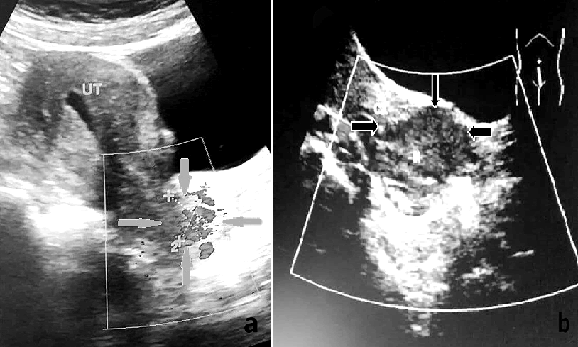
| • Observe the location, state and echo characteristics of cervical cancer lesion and measure the cervical long diameter, anteroposterior diameter and area | ||||
| • Observe the peripheral and internal blood flow of the lesion and record data in time (Figure 1). | ||||
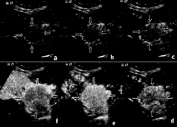
| The process of CEUS examination is as follows: When common two-dimensional ultrasound clearly shows sagittal sections of uteri and cervixes, adjust the instrument to meet imaging conditions; order assistants to draw 2.4 ml SonoVue and perform bolus injection through antecubital veins; all the phases of contrast enhancement were recorded and analyzed. Observe the inflow of contrast agent in the lesion (Figure 2), carefully measure the long diameters, anteroposterior diameters and areas of cervical cancer lesions, and analyze dynamic variation process and enhancement situations of echo intensity at cervical cancer lesions and surrounding tissues. Instrument built-in software automatically generated TIC (time-intensity curve) of cervical cancer lesions and surrounding tissues. Open the continuous dynamic images stored in the instrument, manual draw the outline of ROI (region of interest) in the greatest intensity of the contrast agent in the cervical cancer lesions, measure the long diameter, anteroposterior diameter and area, record data in time. | ||||
| Indicator observation | ||||
| Measure the sizes and areas of cervical cancer lesions and determine the staging of the lesion according to the ultrasound diagnostic criteria through conventional ultrasound and CEUS. The criterion of clinical staging of cervical cancer is according to the Federal International of Gynecology and Obstetrics (FIGO) [3]: Stage 0 (Preinvasive carcinoma or intraepithelial carcinoma, which cannot be detected by ultrasound); Stages I (Carcinoma is limited to the cervix); Stages II (The lesion of carcinoma has exceeded the cervix, but did not reach the pelvic wall); Stages III (Carcinoma invasion of the pelvic wall and the vagina. No gap between tumor and pelvic wall in rectal examination); Stages IV (Carcinoma spread beyond the true pelvis or invasion of the bladder or distant metastasis). The diagnostic criterion of ultrasound in staging of cervical cancer in this study is mainly based on FIGO: Stages I (The lump is located in the cervical muscular layer); Stages II (The lump breaks through the cervix of the uterus, but did not spread to adjacent organs); Stages III (The lump invasion of the pelvic wall and the vagina. Partial patients with hydronephrosis); Stages IV (The lump spread to adjacent organs or distant metastasis). | ||||
| Statistical treatment | ||||
| Statistical analysis was conducted by using the software spss17.0 ± s indicates measurement data; χ2 is used to test measurement data; T test method is used to make inter-group comparison; and P <0.05 indicates statistically significant. | ||||
Results |
||||
| Comparison of sizes and areas of cervical cancer lesions displayed through the two diagnostic methods | ||||
| The cervical cancer lesion area (2328.18 ± 1153.06 mm2), cervical cancer lesion long diameter (51.09 ± 13.25 mm), and cervical cancer lesion anteroposterior diameter (42.56 ± 13.29 mm) displayed through CEUS were significantly better than the cervical cancer lesion area (1667.69 ± 943.34 mm2), cervical cancer lesion long diameter (44.49 ± 13.42 mm), and cervical cancer lesion anteroposterior diameter (36.06 ± 13.47 mm) through conventional ultrasound , which was statistically significant (P <0.05) (Table 1). | ||||
| Comparison of diagnostic results in different stages between the two diagnostic methods | ||||
| The detection rates of stage I cervical cancer (85.18%) and stage II cervical cancer (92.98%) and total detection rate (93.65%) achieved by CEUS were greatly superior to those of stage I (0.00%) and stage II (36.84%) and the total detection rate (50.00%) by conventional ultrasound , which was statistically significant (P <0.05) (Table 2). | ||||
Discussion |
||||
| Cervical cancer is one of the most common gynecological cancers in clinic, which has always been the first place in gynecologic malignant tumor in China. There are many risk factors of cervical cancer, in which the primary one is persistent infection of high-risk HPV, and the second ones are early experience of first sexual life and first delivery, multiple sex partners, repeated pregnancy and delivery, malnutrition and poor health habit, etc. [4]. The patients with early-stage cervical cancer usually have no obvious symptoms and signs, and subsequently are followed by symptoms of colporrhagia and white or bloody fluid. Patients with advanced stage generally have different degrees of secondary symptoms according to lesion-involved regions, which threaten human life seriously [5]. The occurrence and development of cervical cancer follow the principle of from quantitative change to gradual change and finally to sudden change. Once sudden change occurs, it is particularly difficult to start treatment. It can greatly enhance survival rate of patients to detect and confirm cervical cancer and select therapeutic schedule as early as possible. | ||||
| The clinical diagnostic methods of cervical cancer are various such as gynecological examination, ultrasound, CT, MRI and PET, in which ultrasonic examination is more common. In this group of tests, CEUS and conventional ultrasound are respectively used for contrast examinations and preoperative diagnosis for cervical cancer patients. Early diagnosis of cervical cancer can definite the lesion location and surrounding infiltrative situations, which can help the doctor to choose the appropriate treatment plan, controls the tumor proliferation, thus enhances the clinical cure rate, improves the prognosis. However, in early cervical cancer patients, the lesions of cervical are usually very smalland without morphological changes in cervixes, thereforeit is hard to be detected by conventional ultrasound [6]. In contrast, CEUS can clearly show blood perfusion in capillary vessels and microcirculatory changes in tumor lesions, enabling inspectors to observe the sizes and ranges of cervical cancer lesions, the infiltrative situations of surrounding tissues, increasing detection rate of cervical cancer, and then give appropriate treatment based on diagnostic results [7]. Early cervical cancer is clinically manifested as cervical fester. Conventional ultrasound can detect enlarged cervix, irregular morphous, and substantial heterogeneous low-echo mass in the inner part, but cannot clearly show infiltrative situations of cervical cancer lesionsbecause there is no clear boundary between endometrium and myometrium at lower uterine segment. In this case, it is difficult for medical staff to make judgement according to the examination results. Only when the cervical morphology has a great advance can a part of stage I cervical cancer patients are confirmed. The value of conventional ultrasound in the diagnosis of early cervical cancer is low. CEUS is easy to operate and can be used repeatedly without time limit and radiation. CEUS has extremely high resolution and strong sensibility, thereforeit can clearly show fibrous layer of cervical inner lining, depth of lesion infiltration in myometrium, and changes of lymphoid tissue around cervix and cervical mucosa, and can detect heterogeneous enhancement in the inner part of lesions. It effectively enhances diagnostic sensitivity and specificity and has a high value in preoperative and staging diagnosis [8]. According to the results of present study as shown in Table 1, the cervical cancer lesion area, cervical cancer lesion long diameter, and cervical cancer lesion anteroposterior diameter displayed through CEUS were significantly better than those of conventional ultrasound , which was statistically significant (P<0.05). This indicates that compared with conventional ultrasound , CEUS has higher sensibility and resolution, and can clearly show the area, long diameter and anteroposterior diameter of cervical cancer lesion, enabling inspectors to accurately locate lesions and determine their infiltrative ranges. CEUS provides objective imaging materials for clinical diagnosis, which are helpful for selecting appropriate therapeutic schedule, so as to reduce death rate and improve survival quality of patients [9]. According to the result of this study as shown in Table 2, the detection rates of stage I and stage II cervical cancer and total detection rate achieved by using CEUS were all greatly superior to those of conventional ultrasound, which was statistically significant (P<0.05), while the comparison of detection rates of stage III and stage IV cervical cancer between CEUS and conventional ultrasound was statistically insignificant (P>0.05). As the lesions of early cervical cancer patients are small, conventional ultrasound cannot effectively show the situations of inner part of lesions and neighbouring areas, but the sizes and infiltrative ranges of lesions achieved by CEUS were obviously larger than those of conventional ultrasound. This indicates that CEUS can detect concrete situations of cervical cancer to a large extent, with higher detection rates and more accurate examination results. CEUS is more favourable than conventional ultrasound in preoperative and staging diagnosis, for it has a high application value in provide more effective basis for selecting clinical therapeutic schedule [10]. | ||||
Conclusion |
||||
| In conclusion, compared with conventional ultrasound, CEUS, due to it higher resolution and sensibility, can clearly display the boundary of cervical cancer lesions and surrounding infiltrative situations; it has higher detection rate and provides reliable basis for selection of clinical therapeutic schedule. In light of its high application value, it is recommended to promote the use of CEUS. The main focus of this study is the ability of the two kinds of methods in detecting and staging of cervical cancer. Therefore, there is not pay more attention on whether the size of the ultrasonic measurement is consistent with the size of the surgical resection, it should pending further analysis and research. | ||||
Tables at a glance |
||||
|
||||
Figures at a glance |
||||
|
||||
References |
||||
|

