ISSN: 0970-938X (Print) | 0976-1683 (Electronic)
Biomedical Research
An International Journal of Medical Sciences
Research Article - Biomedical Research (2016) Volume 27, Issue 4
Characterization of viscoelastic behavior of rat cervix in the last trimester of pregnancy
Alireza Ashofteh Yazdi1*, Ali Esteki2, Mohammad Mehdi Dehghan3 and Farhad Tabatabai Ghomsheh4
1Department of Biomedical Engineering, Science and Research Branch, Islamic Azad University, Tehran, Iran
2Department of Biomedical Engineering and Physics, Shahid Beheshti University of Medical Sciences, Tehran, Iran
3Department of Surgery & Radiology, Faculty of Veterinary Medicine, University of Tehran, Tehran, Iran
4Department of Ergonomics, University of Social Welfare and Rehabilitation Sciences, Tehran, Iran
- *Corresponding Author:
- Alireza Ashofteh Yazdi
Department of Biomedical Engineering, Science and Research Branch, Islamic Azad University, Tehran, Iran
Accepted April 11, 2016
Quantitative measurement of mechanical properties of cervix is an effective tool to diagnose cervical insufficiency, specifically in the last trimester of pregnancy when preterm birth is mostly probable to occur. The objective of this study was to characterize the viscoelastic behavior of rat cervix at different displacement levels and rates early in the last trimester of pregnancy. Distinct displacement levels and rates were employed to measure the tensile and load-relaxation properties of cervices from 16 days postconception pregnant rats. After preconditioning, 2 millimeters uniaxial displacement was applied to the distal halves of five rat’s cervices circumferentially at displacement rates of 0.01, 0.1 and 1 mm/sec. This displacement was held for 10 minutes while the tissues were mostly relaxed. The same procedure was repeated for the displacement levels of 1, 1.5 and 2 millimeters at the rate of 1 mm/sec. Tensile and loadrelaxation curves were well described by a quasi-linear viscoelastic model. Statistical analysis revealed significant correlation between the change in displacement levels and rates, and the elastic response of the tissues. The viscous response, though, did not show strong dependency on displacement levels as well as displacement rates. Cervix becomes considerably stiffer and exhibits less viscous effects in increasing rates. However, higher displacement levels result in lower strength of the tissue. These results show the viscoelastic behavior of cervix in the last trimester of pregnancy. These mechanical characterizations may be useful for prediction of cervical insufficiency and preterm birth
Keywords
Cervical insufficiency, Preterm birth, Viscoelastic behavior, Displacement levels and rates.
Introduction
Characterization of viscoelastic behavior of cervix will provide insight to the changes of elastic and viscous responses of the tissue during pregnancy. This detailed knowledge will help us understand the effect of clinical and physiological variables on the normal functional behavior of the pregnant tissue. The cervix is a mechanical barrier that holds the growing fetus inside and stays firm until the fetus is mature. At the time of delivery, the cervical tissue softens and ripens to allow the baby to pass [1]. Cervical Insufficiency refers to a condition in which cervix dilates asymptomatically in the absence of contractions from the uterine smooth muscles and results in a spontaneous pregnancy loss between the second and third trimester of pregnancy. Cervical Insufficiency accounts for a significant percentage of extremely premature deliveries with high incidence of infant mortality [2].
Changes in mechanical properties of cervix during pregnancy, is a result of microstructural and compositional changes happen as gestational age increases. These changes include increase in the tissue hydration, collagen extractability and sulfated glycosaminoglycan content but the amount of collagen content remains the same [3,4]. These changes may cause the tissue to lose its mechanical properties, especially in the last trimester of pregnancy, and becomes insufficient to retain the fetus until term. In this case preterm birth is most likely to happen [5]. Characterizing the material behavior of human cervical tissue using either in vivo or ex vivo methodologies remains a challenge. Each methodology has advantages and drawbacks in terms of its ability to capture tissue remodeling characteristics of pregnant tissue or to obtain enough data to derive fully predictive 3D constitutive material equations. Animal models of pregnancy offer opportunities to test gestation timed, genetically-altered, or chemically-treated samples. However, it is unclear whether the factors driving tissue remodeling in these animal models are similar enough to humans [6].
Ex vivo mechanical tests allow for control over definitions of boundary and loading conditions such that careful analysis of the tissue’s stress response to various loading scenarios can be assessed. A large range of variation reported for human cervical tissue tangent moduli which are due to the variation in testing protocols, tissue age and parity between the studies [7,8]. In vivo interrogation of the cervix during pregnancy allows characterization of longitudinal changes over the course of the gestation. The driving motivation for the development of the in vivo tools is both investigational and diagnostic in nature, where the ultimate clinical goal is to uncover cervical biomarkers that are predictive of preterm birth [8].
Bishop score is a primary method used via a digital examination of the cervix, which measures both structural and material parameters such as effacement, dilation and softness [9]. These measurements can be highly variable due to the subjective nature of each component and lack of precision. Transvaginal ultrasound techniques have been popularly used recently to measure anatomical parameters of cervix, such as changes in the length of the tissue during pregnancy [10,11]. Although anatomical parameters are useful indicators of preterm birth, but these measurements alone have low predictive value and in many cases did not outperform the bishop score in predicting preterm birth [12,13]. Ultrasound elastography is a method recently used in the field of gynecology. Semi-quantitative elastography can be employed to evaluate changes in cervical stiffness during pregnancy. This method estimates the average tissue displacement (strain) within a defined region of interest when oscillatory compression is applied. However, this method is a good predictor of cervical function but before it can be used, the mechanical properties of tissue must be characterized [14].
Due to the limitations of human cervical tissue sampling, rodent models have been widely used to investigate for understanding physiological changes of the cervix during pregnancy. Despite the differences in the mechanical properties of the rat cervix to the humans, the timing and degree of change makes it as a good candidate for understanding events that precede preterm birth [15,16]. Table 1 summarizes uniaxial tests results reported in the literature for the rat cervical tissue. Each study measured the QLV model constants at defined displacement levels and rates [4,16].
| References | Specimen description | Strain rate & Strain level | Tangent modulus (MPa) | QLV model constants |
|---|---|---|---|---|
| W. Barone et al., 2010 | NP, MP and PP Term, Long Evans rat cervix Rectangular specimen, Compression test | Rate: 0.167 mm/s Level: 0.2 | Not given | B ex. os NP:38.8 |
| B ex. os MP:53.4 | ||||
| B ex. os PP:39.9 | ||||
| D ex. os NP:0.124 | ||||
| D ex. os MP:0.143 | ||||
| D ex. os PP:0.148 | ||||
| MJ. Poellmann et al., 2012 | PG Term (15-21 days pregnancy), Sprague-Dawley rat cervix, Tensile test | Rate: 0.02 mm/s Level: 0.25 | K Day 15: 66.21 g/mm | B ex. os Day 15: 2.3 |
| K Day 21:7.34 g/mm | B ex. os Day 21: 1.6 |
Table 1. Rat cervical tissue tangent moduli and QLV model constants from uniaxial ex vivo mechanical tests. The strain level reported is location of the tangent modulus. PG: Pregnant, NP: Non-Pregnant, MP: Mid-Pregnant, PP: Post-Partum, ex: External.
Accurate diagnostic criteria and treatment guidelines for prediction of preterm birth in case of cervical insufficiency are not established and remains a clinical and research challenge. Quantitative mechanical diagnostic techniques have been gaining in popularity in diagnosis of cervical insufficiency [17]. Therefore the objective of this study was to characterize the viscoelastic response of rat cervix at different displacement levels and rates in the last trimester of pregnancy. These characterizations may enhance the development of medical devices which measure the viscoelastic response of human cervix to defined applied variables for diagnosis of the abnormal behavior of tissues.
Materials and Methods
Tissue collection
All procedures were approved by the institutional animal care and use committee at the University of Tehran. The samples were obtained from the Animal Lab of the Veterinary Surgery Department of the University of Tehran. Twenty Virgin Wistar rats with the same characteristics (age, weight) were kept in five different boxes and were mated by male ones at a definite time. After a night males removed and five rats were found to be pregnant. After euthanasia with carbon dioxide, cervices were harvested from 16 days post-conception pregnant rats early in the last trimester of pregnancy (full term ̴ 22 days). The cervix was excised from the uterus at the transition zone. Then each cervix was cut in half perpendicular to the cervical canal, as illustrated in Figure 1a. Cervical mucus was not removed prior to testing to prevent damage resulting from its removal. The distal halves were kept in normal saline and taken immediately to the laboratory for mechanical testing to avoid tissue damage or change of in-vivo mechanical properties. The proximal portion has been shown to have higher smooth muscle and less collagen content compared to the distal portion [16]. Table 2 presents the information on collected specimens.

Figure 1A. Illustration of the rat cervix. Cervix was excised from the uterus at the transition zone. Then each cervix was cut in half perpendicular to the cervical canal. The distal halves were kept in normal saline and taken immediately to the laboratory for mechanical testing. The rat cervix has two canals one leading to each uterine horn. For mechanical testing, stainless steel rods were threaded through each canal.
| No. | Width (mm) | Length (mm) | Thickness (mm) |
|---|---|---|---|
| 1 | 5.5 | 5 | 1.1 |
| 2 | 5.5 | 4 | 1.1 |
| 3 | 5.1 | 5 | 1.1 |
| 4 | 5.4 | 4.3 | 1.1 |
| 5 | 5.6 | 4.1 | 1.1 |
Table 2. Sample information (Cervices samples from Wistar rats of age 7 months with 250 ± 10 grams weight)
Mechanical testing
The length, width and thickness of the each half were measured using digital calipers with an accuracy of ± 0.01. A pair of steel rods each 1 mm diameter, were threaded through the cervical canals. The rods were placed into a custom-made fixture, as shown in Figure 1b. Once the cervix was placed in the fixture, the distance between the rods was measured to ensure that the dimensions were not changed by the calipers due to deformation. All tensile tests were performed by a uniaxial test device, designed and fabricated to measure mechanical properties of soft biological tissues [18]. The testing device is equipped with a 10-N load cell with an accuracy of ± 0.5 %.

Figure 1B. Cervix positioned in the custom designed fixture attached to the uniaxial tensile test device, designed and fabricated to measure mechanical properties of soft biological tissues.
Displacement controlled tensile loadings were implemented to the samples circumferentially in medial-lateral direction. In order to obtain a repeatable mechanical behavior, tissues were preconditioned. For this purpose, samples underwent 10 cycles of loading-unloading at low displacement levels at the rate of 1 mm/sec to coincide stress-strain curves. After preconditioning, the displacement of 2 millimeters was applied to the samples circumferentially. The displacement rates of 0.01, 0.1 and 1 mm/sec were employed to carry out the tension tests at the ramp section. They defined as low rate, mid-rate and high rate, respectively. Furthermore, Samples were stretched to the displacement levels of 1, 1.5 and 2 millimeters at the rate of 1 mm/sec to investigate the effect of displacement levels on the viscoelastic response of the tissue. A recovery period of 5 minutes was considered between tests. To study the relaxation behavior of tissues, the displacement was held for 10 minutes while the tissues were mostly relaxed, as illustrated in Figure 1c. Samples were sprayed with saline solution to prevent drying of the tissue during the experiments. Force, displacement and time were collected at 30 Hz.

Figure 1C. Samples were stretched at different displacement rates to different displacement levels in the ramp section, and then kept at these levels of displacement for 10 min during the hold section.
Mechanical analysis
Stress and strain were calculated using the force and displacement measured in the tests as functions of time. Curve fitting tool in Matlab was used to fit best quasi-linear viscoelastic (QLV) models to the stress-strain and stress-time curves using least square method. To analyze material properties of cervix, stress was calculated by normalizing load by the cross-sectional area and strain was defined as tissue displacement divided by initial internal width. A rectangular geometry was assumed for all samples [16].
Elastic modulus (E) was defined as the slope of the linear region of stress-strain curves at the end of each ramp and defined in MPa [19]. The tensile and load-relaxation data were fit to a quasi-linear viscoelastic (QLV) model [20]. The quasilinear viscoelastic (QLV) model parameters were extracted to compare the viscoelastic properties of samples at different displacement levels and rates. A coefficient of determination R2>0.9 was considered for the acceptance of the best model fitted.
The instantaneous elastic response of the tissues was described by the QLV model as a function of strain (Equation 1):
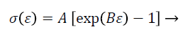 (1)
(1)
Where parameter B indicates the rate of change in the slope of the stress-strain curves. The relaxation behavior of tissues was well described by a four-parameter function which closely fit the stress of the hold section as a function of time (Equation 2):
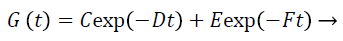 (2)
(2)
Where parameters D and F indicate the slope of the short term and long term relaxation of the stress-time curves.
Statistical analysis
Statistical analysis was performed using SPSS statistics 18 software. One way ANOVA was used to study the significance of the independent variables namely, displacement levels and rates, on measured viscoelastic properties of the samples. A Pvalue of less than 0.05 was considered as significant statistical difference. This limit can be extended due to the diversity of the viscoelastic response of the tissue. Furthermore, a Duncan post-hoc test was carried out to classify the results at different displacement levels and rates. The estimated QLV parameters were represented graphically with box plots.
Results
Elastic response
The uniaxial tension tests performed on 5 samples from cervices of 16 days post-conception pregnant rats. Each sample was tested at three different displacement levels and rates. Results revealed that the elastic modulus slightly increased at high rate compared to low rate and mid-rate, as illustrated in Figure 2a (p=0.215). The difference between the elastic modulus was found significant particularly at high rate and low rate (p=0.024). Though, the elastic modulus did not change considerably in increasing displacement levels, as shown in Figure 2b (p=0.744).

Figure 2A. The elastic modulus slightly increased as displacement rate increased (p = 0.215).

Figure 2B. The elastic modulus did not change considerably in increasing displacement levels (p = 0.744).
Parameter B, which is the rate of change in the slope of the elastic response and governs the nonlinearity of the stressstrain curve, was found to increase significantly as rate increased, as illustrated in Figure 2c (p=0.000). A Duncan post-hoc test showed that the mean B values of tissues at low rate and mid-rate were in the same subset while it increased considerably at high rate. In addition, Parameter B showed considerable dependency on displacement levels and decreased significantly in increasing levels (p = 0.029). A Duncan posthoc test showed that the mean B values of tissues at displacement levels of 1.5 and 2 millimeters were in the same subset while it was considerably higher at the level of 1 millimeter (Figure 2d).

Figure 2C. The rate of change in the slope of the elastic response significantly increased at higher displacement rates (p = 0.000).

Figure 2D. The rate of change in the slope of the elastic response significantly decreased in increasing displacement levels (p = 0.029).
Viscous response
The uniaxial tension test was held at the defined displacement level for 10 minutes while the tissues were mostly relaxed. Parameter D, which represents the initial slope of the relaxation curve, slightly increased at higher displacement rates, as illustrated in Figure 3a (p=0.219). It was found that D value differs considerably at high rate and low rate (p=0.07). In addition, no significant difference was seen for parameter D in increasing displacement levels, as shown in Figure 3b (p=0.421).

Figure 3A. The initial slope of the relaxation curve slightly increased as displacement rate increased (p = 0.219).

Figure 3B. The initial slope of the relaxation curve did not change considerably as displacement level increased (p=0.421).
For parameter F, which represents the late slope of the relaxation curve, no difference was detected as displacement rate increased, as illustrated in Figure 3c (p = 0.58). Furthermore, higher displacement levels did not result in change of parameter F, as shown in Figure 3d (p=0.99).

Figure 3C. The late slope of the relaxation curve did not change considerably at higher displacement rates (p=0.58).

Figure 3D. The late slope of the relaxation curve did not change considerably in increasing displacement levels (p = 0.99).
Discussion
Quantification of viscoelastic properties of rat cervix in the last trimester of pregnancy has been done in previous studies [4,16]. Nevertheless, this was the first work to study the effect of different displacement levels and rates on viscoelastic properties of rat cervix in late stage of pregnancy. Additionally, unlike some previous studies based on structural properties of the tissue, the intrinsic material properties of cervix were estimated which are independent of the sample geometry and size. The results of our study strongly proved that the pregnant tissue is rate dependent and its viscoelastic properties change at different displacement levels and rates.
The critical parameters estimated by tensile and load-relaxation curves utilizing QLV theory were the rate of change in the slope of the elastic response B, and the initial and late slope of the relaxation curve, D and F. The elastic modulus and parameter B, associated with the elastic response of cervix, increased as displacement rate increased. Though, the parameter B significantly decreased in increasing displacement levels. These changes show that by increasing the displacement rate tissue becomes stiffer and its nonlinearity increases as well, while tissue’s softening behavior occurs in increasing displacement levels. It was found that increasing the displacement rates result in increase of the initial slope of the relaxation curves. The late slope of the relaxation curves did not change in increasing displacement levels and rates. As Equation 3 defines, the time constant of the relaxation curve (τ) is equal to the viscous parameter (η) divided by the elastic modulus (E) of the tissue. What can be found is that by increasing the displacement rate, pregnant tissues relax faster and the time at which relaxation terminates reduces which is due to less viscous parameter affecting.
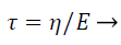 (3)
(3)
The increased strength of cervix at higher displacement rates is desirable, specifically in the beginning of the last trimester of pregnancy when preterm birth is mostly probable to occur in case of cervical insufficiency. The softening behavior of the pregnant tissue at higher levels of displacement reduces its resistance to abrupt loads early in the last trimester of pregnancy. This strength reduction can be a major cause of spontaneous preterm birth while higher physiological strains are inserted to the cervix. Additionally, less viscous effect enables the tissue to recover faster to its steady state. This fast recovery allows for loads to be dissipated more quickly. These changes may enhance the ability of the cervix to maintain load bearing capacity and efficient absorption of the loads. These changes are related to the altered matrix organization and porosity of the pregnant tissue reported by others [3,21].
Consequently, increase of tissue’s strength in increasing displacement rates and its decrease in increasing displacement levels is probable for the pregnant cervix. Furthermore, decrease of the viscous effects in increasing displacement rates enhances the dissipation ability and load absorption of the tissue. These changes in mechanical properties may provide criteria for the normal viscoelastic behavior of the pregnant cervix. Tissues not responding to the applied variables with such a trend are known as abnormal and predicted to have the potential of cervical insufficiency early in the last trimester of pregnancy. This hypothesis must be further studied clinically on human cases to be widely approved.
There are some limitations of this study to be considered for the future works. Despite cervical insufficiency and preterm birth occurs early in the last trimester of pregnancy, more time points are needed to measure the viscoelastic properties of cervix at distinct displacement levels and rates. The second limitation is that mechanical testing is limited to static loading, so the static viscoelastic properties were obtained. Dynamic mechanical analysis is essential to know the dependency of the complex viscoelastic properties of cervix to the desired variables. Assuming rectangular geometry for the tissue was used for the simplicity to estimate material properties of the samples. This assumption may affect the mechanical test results estimated.
Conclusions
The goal of this work was to characterize the viscoelastic behavior of rat cervix at different displacement levels and rates early in the last trimester of pregnancy. The tensile and loadrelaxation parameters change as displacement levels and rates increase. Cervix becomes considerably stiffer and exhibits less viscous effects in increasing rates. However, higher displacement levels result in lower strength of the tissue. These results show the viscoelastic behavior of cervix in the last trimester of pregnancy. These mechanical characterizations may be useful for prediction of cervical insufficiency and preterm birth.
Ethical Approval
All applicable international, national and institutional guidelines for the care and use of animals were followed.
References
- Babacan O. Examining viscoelastic response of pregnant rat cervical tissue using 3, 4 and 5 element spring and dashpot models. Master thesis University of Illinois at Urbana-Champaign 2012.
- Paskaleva AP. Biomechanics of cervical function in pregnancy- case of cervical insufficiency. Doctoral Dissertation Massachusetts Institute of Technology 2007.
- Myers KM, Paskaleva AP, House M, Socrate S. Mechanical and biochemical properties of human cervical tissue. ActaBiomaterialia 2008; 4: 104-116.
- Poellmann MJ, Chien EK, McFarlin BL, Wagoner Johnson AJ. Mechanical and structural changes of the rat cervix in late-stage pregnancy. Journal of the Mechanical Behavior of Biomedical Materials 2013; 17: 66-75.
- House M, Socrate S. The cervix as a biomechanical structure. Ultrasound in Obstetrics and Gynecology 2006; 28: 745-749.
- Elovitz M, Mrinalini C. Animal models of preterm birth. Trends in Endocrinology and Metabolism 2004; 15: 479-487.
- Yao W, Yoshida K, Fernandez M, Vink J, Wapner R, Ananth C, Oyen M, Myers K. Measuring the compressive viscoelastic mechanical properties of human cervical tissue using indentation. Journal of the Mechanical Behavior of Biomedical Materials 2014; 34: 18-26.
- Myers KM, Feltovich H, Mazza E, Vink J, Bajka M, Wapner RJ, Hall TJ, House M. The mechanical role of the cervix in pregnancy. Journal of Biomechanics 2015.
- Bishop EH. Pelvic scoring for elective induction. Obstetetrics and Gynecology 1964; 24: 266-268.
- Crane JM, Hutchens D. Use of transvaginal ultrasonography to predict preterm birth in women with a history of preterm birth. Ultrasound in Obstetrics and Gynecology 2008; 32: 640-645.
- Gramellini D, Fieni S, Kaihura C, Modena AB. Cervical length as a predictor of preterm delivery: gestational age-related percentiles vs fixed cut-offs. ActaBiomedica 2007; 78: 220-224.
- Iams JD. Prediction and early detection of preterm labor. Obstetrics and Gynecology 2003; 101: 402-412.
- Newman RB, Goldenberg RL, Iams JD, Meis PJ, Mercer BM, Moawad AH, Thom E, Miodovnic M, Caritis SN, Dombrowski M, Thurnau GR. National institute of child health and human development maternal-fetal medicine units’ network, preterm prediction study: comparison of the cervical score and Bishop score for prediction of spontaneous preterm delivery. Obstetrics and Gynecology 2008; 112: 508-515.
- Hernandez-Andrade E, Hassan SS, Ahn H, Korzeniewski SJ, Yeo L, Chaiworapongsa T, Romero R. Evaluation of cervical stiffness during pregnancy using semiquantitative ultrasound elastography. Ultrasound in Obstetrics and Gynecology 2013; 41: 152-161.
- Mahendroo M. Cervical remodeling in term and preterm birth: insights from an animal model. Reproduction 2012; 143: 429-438.
- Barone WR, Feola AJ, Moalli PA, Abramowitch SD. The effect of pregnancy and postpartum recovery on the viscoelastic behavior of the rat cervix. Journal of Mechanics in Medicine and Biology 2012; 12: 1250009-1250026.
- Dewall RJ, Varghese T, Kliewer MA, Harter JM, Hartenbach EM. Nonlinear mechanical behavior of cervical tissue with increasing pre-compression. In: Proc. 2009 IEEE International Ultrasonics Symposium; 2009.
- Faturechi R, Hashemi A, Fatouraee N. Do mechanical properties of human fetal membrane depend on strain rate?. Journal of Obstetrics and Gynecology Research 2014; 41: 84-92.
- Sepehri B, Taheri E, Ganji R. Biomechanical analysis of diversified screw arrangement on 11 holes locking compression plate considering time-varying properties of callus. Biocybernetics and Biomedical Engineering 2014; 34(4): 220-229.
- Nekouzadeh A, Pryse KM, Elson EL, Genin GM. A simplified approach to quasi-linear viscoelastic modeling. Journal of Biomechanics 2007; 40: 3070-3078.
- Mazza E, Nava A, Bauer M, Winter R, Bajka M, Holzapfel GA. Mechanical properties of the human uterine cervix: an in vivo study. Medical Image Analysis 2006; 10: 125-136.