ISSN: 0970-938X (Print) | 0976-1683 (Electronic)
Biomedical Research
An International Journal of Medical Sciences
- Biomedical Research (2015) Volume 26, Issue 3
Biochemical and molecular characterization of cerebrospinal fluid for the early and accurate diagnosis of Mycobacterium tuberculosis.
Sunita D Singh, Tariq Masood, Ravjit K Sabharwal, Narotam Sharma, Satish C Nautiyal, R.K. Singh
Department of Biochemistry, Central Molecular Research Laboratory, SGRR Institute of Medical & Health Sciences, Dehradun 248001, India
- Corresponding Author:
- R K Singh
Department of Biochemistry
Shri Guru Ram Rai Institute of Medical
and Health Sciences, Patel Nagar
Dehradun 248001(Uttarakhand)
India
Accepted date: March 25 2015
The present study includes 50 patients symptomatic for tuberculous meningitis and 15 patients with meningitis other than tuberculosis The CSF specimens were subjected for ADA titer estimation, AFB smear preparation and molecular characterization by Nested-PCR for IS6110 gene. Out of 50 specimens 2 came AFB smear positive whereas 29 specimens came positive for Tuberculosis (TB) PCR and 21were negative for TB PCR. 17 specimens came positive by both the targets IS6110 and mpb-64 gene but 9 specimens came positive only by IS6110 gene, but negative for mpb- 64 gene. 3 specimens came positive for mpb-64 gene and the same were negative for IS6110 gene. 29 specimens came positive for Multiplex PCR. ADA activity >10 (U/L) was found in 33 specimens. The positive signals obtained in these CSF specimens were higher with IS6110 system 26/50 (52%) than with mpb-64 system 20/50 (40%). PCR gave a sensitivity of (58%) and specificity of (93.3%), Positive predictive value of (96.6%) and Negative predictive value of (40%) for the diagnosis of TBM in 50 patients who were strongly suspected for Tuberculosis meningitis. ADA activity in the CSF had limited value when using a cut-off value of 10 U/L for the diagnosis of tuberculous meningitis. In actual clinical practice, the diagnosis of CNS tuberculosis requires not a screening examination but rather a definitive molecular procedure like IS6110 and mpb-64 gene characterization for accurate and confirm diagnosis of tubercular meningitis.
Keywords
Tuberculous meningitis, Conventional PCR, Anti-tuberculosis drugs, Adenosine deaminase Activity, Antituberculous therapy
Introduction
Tuberculosis (TB) is an infectious bacterial disease caused by Mycobacterium tuberculosis, typically affects the lungs, but it can also affect other parts of the body. The World Health Organization has noted that the global incidence of TB is increasing by 0.4% per annum [1, 2]. Tuberculosis can be pulmonary and extra-pulmonary and Central nervous system (CNS) TB, accounts for about 5% of all extra-pulmonary TB and tuberculosis meningitis (TBM) is the most serious complication. Delay in the diagnosis and institution of therapy can result in death or severe neurological defects. In TBM, accurate and rapid diagnosis and early treatment for tuberculosis are the most important factors with regard to the prognosis and the prevention of long-term neurological sequel [3]. A diagnostics method includes serological, radiological and microbiological investigations. However, the “gold standard” method based on culturing the M.Tb, including the direct smear examination for acid-fast bacilli (AFB), is inadequate for early diagnosis of pauci-bacilliary TB especially TBM, owing to the poor sensitivity or the long time required (4–8 weeks) for cultures [4]. Nucleic acid amplification techniques (NAAT) such as polymerase chain reaction (PCR) have been reported to be more sensitive and specific. Several M. tuberculosis specific sequences such as IS6110, Protein antigen b (13), mpb-64and 65kDa have been evaluated [5, 6]. The present study was planned to focus on the comparative analysis of different assays i.e. ADA titer and Nested-PCR targeting IS6110, Conventional- PCR targeting mpb-64and multiplexing of the mpb-64and IS6110 genes. Conventional and N-PCR for the detection of Mycobacterium DNA sequences in CSF specimens were studied and the DNA sequences were further compared with the ADA levels for the better management of TBM patients.
Materials and methods
Specimen collection
The CSF specimens were collected from the Neurosurgery, Medicine and the Pediatrics Departments of Shri Mahant Indiresh Hospital, Dehradun. This study was approved by Institution ethical clearance committee and the written con sent from all the patients were obtained. Fifty tuberculous meningitis patients with sub-acute or chronic fever with features of meningeal irritation such as headache, neck stiffness and vomiting with or without other features of CNS involvement and 15 non- Tuberculous meningitis patients with high fever and/or signs of meningeal irritation , or head injury and who had received broad-spectrum antibiotics, or with or without CNS manifestations and CSF findings showing increased proteins, decreased glucose (CSF: blood glucose ratio < 0.2), and/or pleocytosis with a predominance of polymorphonuclear cells and good clinical response to broad-spectrum antibiotics were included in this study.
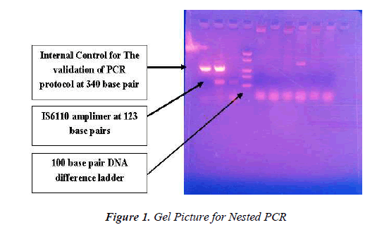
All the CSF specimens were subjected for ADA estimation and amplification of the targets, mpb-64, IS6110 conventional, nested and multiplex PCR. All the CSF specimens were subjected parallel for MTB complex detection by nested PCR using uracil-N-glycosylase (UNG) enzyme in pre-mix targeting IS6110 by conventional PCR using mpb- 64 gene for MTB complex detection and multiplex PCR by IS6110 and mpb 64. All three protocols were subjected with controlled parameters utilizing nuclease free water as negative control where after every three specimens a negative control was processed to check any sort of contamination. Nested PCR was performed utilizing manufacturer protocol (Bangalore genei). In case of nested PCR, an amplification product of size 123 bp was indicative of infection with Mycobacterium tuberculosis complex where as the amplification product of internal control DNA was 340 bp. In case of conventional PCR only 123 base pair product indicates Mycobacterial infection as depicted in figure1 [6, 7].
Results
IS6110 and mpb-64 gene sequences in the M. tuberculosis complex was used to analyze CSF in TBM patients. The collected CSF specimens were analyzed for AFB smear, ADA titre estimation. DNA from 50 cases was isolated by Silica column adsorption method and further used as template individually for IS6110 gene and mpb-64 PCR (Table-2). The same DNA was used for the standard and optimization of multiplex PCR assay develop which included both the genes i.e. IS6110 and mpb-64. It was seen that out of 50 AFB smear patients only 02 came positive. Out of 50 specimens which were subjected separately for IS6110 gene and mpb-64gene both PCR, it was found that 29(58%) came positive where 21 (42%) were negative (Table 2 and 3). Further 17 cases came positive by both the PCR (Table-4). IS6110 gene got amplified alone in 9 (18%) cases, where in 03 (6%) case mpb-64 gene got amplified (Table 3). It was also demonstrated that in-house developed PCR assays targeting IS6110 gene and mpb- 64 gene also got the same result as that with include Nested-PCR and Conventional-PCR (Table-5).
The ADA titre estimation showed the range in between 2.6-146.4 (U/L) (Table-4) was showed in other studies the ADA activity is increased in various other diseases. Hence, the ADA activity doesn’t give a confirmed report of positive tuberculosis meningitis. ADA activity in the CSF had unlimited value when using a cut-off value of 10 U/L for the diagnosis of tuberculous meningitis. The ADA determination in CSF has limited utility for the diagnosis of tuberculous meningitis patients, the major problem was lacking of specificity. Thus, CSF ADA activity is not recommended as a routine diagnostic test for TB of the central nervous system. In an attempt to increase diagnostic accuracy, we performed multiplex PCR using two targets, i.e. IS6110 and mpb-64, together for diagnosis of Mycobacterium tuberculosis complex. We were able to pick up those cases which were missed by IS6110 and mpb-64, as the use of two targets together increased the sensitivity of NAA test. PCR gave a sensitivity of (58%), specificity of (93.3%), Positive predictive value of (96.6%) and Negative predictive value of (40%) for the diagnosis of TBM in patients for strongly suspected for Tuberculosis meningitis.
Discussion
Tubercular meningitis (TBM) which occurs in 7–12% of tuberculosis patients in developing countries like India involves the central nervous system (CNS) and is one of the most severe forms of extra-pulmonary tuberculosis [8,9]. The rapid detection of the causative organism (Mycobacterium tubercular Bacilli) is of paramount importance in TBM as the disease can be fatal and clinical outcome depends heavily on the stage at which treatment is initiated. Diagnosis of TBM is presumptive and is based on clinical symptoms, neurological signs, cerebrospinal fluid (CSF) findings, CT scans and the response to anti-tuberculosis drugs. At present, the diagnosis of CNS tuberculosis remains a complex issue because the most widely used conventional “gold standard” based on bacteriological detection methods, such as direct smear and culture identification, cannot rapidly detect Mycobacterium tuberculosis in CSF specimens with sufficient sensitivity in the acute phase of TBM. Rapid techniques based on nucleic acid amplification such as PCR are more sensitive and specific as they attempt to detect specific DNA sequences of the organism [10]. In addition, the innovation of nested PCR assay technique is worthy of note given its contribution to improve the diagnosis of CNS tuberculosis. Currently, conventional qualitative analysis assay makes it possible to perform accurate quantitative analyses with a high degree of reproducibility [11].
The use of molecular biology techniques in the diagnosis of tuberculosis started with the use of DNA probes (Grange, 1989), which were less sensitive than even the existing conventional tests. They have been increasingly used for this purpose since the introduction of the PCR technique. The majority of the investigators performing PCR-based diagnosis of tuberculous meningitis have used insertion sequence IS6110 as a target [12]. The principal reason for using IS6110 is the presence of multiple copies in the M. tuberculosis genome which was thought to confer higher sensitivity. It has, however, been shown that there are M. tuberculosis strains originating from India which do not contain IS6110. Acid Fast Bacilli (AFB) staining is a common, rapid and specific method for M. tuberculosis detection, but in case of Tuberculosis meningitis (TBM), AFB results are mostly negative because the bacterial load in the CSF sample is very low as the cell doubling time of a Mycobacterium tuberculosis is around 16 to 18 hours which is much higher than a normal bacterial cell doubling time [10]. However, if culture of bacterial cells is performed then it can give the positive results. Adenosine deaminase Activity (ADA) estimation in CSF is not only simple, inexpensive and rapid but also fairly specific method. The results of this study indicated that ADA determination in CSF has limited utility for the diagnosis of tuberculous meningitis patients [11]. The major problem was lacking of specificity. ADA is a useful surrogate marker for TB because it can be detected in body fluids such as pleural, pericardial and peritoneal fluid. The levels of ADA increase in TB because of the stimulation of T cells by mycobacterial antigens [12-15].
Increased ADA levels are found in various forms of Tuberculosis. Hence, it is a marker for tuberculosis. However, ADA activity is increased in various other diseases like mononucleosis, typhoid, viral hepatitis, initial stages of AIDS and in malignant tumors. Hence, the ADA activity doesn’t give a confirmed report of positive tuberculosis meningitis. However, if it is followed with TB PCR then it can give the positive results. Hence, the ADA activity should be followed with TB PCR. As nested PCR itself increases the sensitivity and specificity of an assay and when Uracil N Glycosylase (UNG) is being utilized in pre-mix will make the assay a significant molecular diagnostic tool for Mycobacterium tuberculosis complex detection. Nested PCR for tuberculosis detection is a better tool when incorporated with an addition of UNG to prevent amplicon contamination. False positive cases by amplicon contamination can be prevented by UNG and dUTP instead of dTTP. The skill set required to adequately treat critically ill patients will also require knowledge of molecular biology for better diagnosis and treatment.
The foundations of molecular biology and genetics are essential for the understanding of the mechanisms of disease. Correct, novel, significant molecular diagnostic tools are very important for all those laboratories performing routine diagnosis of tuberculosis in PCR based laboratories settings. In addition, particular emphasis should be applied to quality control and quality assurance programs in clinical laboratories which employ any new diagnostic approaches. Amplicon contamination detection and its prevention is of critical importance where the results interpretations are directly involved with patient’s health. Acceptance and implementation of PCR in the diagnostic laboratory requires an understanding of its mechanics, meaning of results, the test’s limitations, and being able to recognize superior to various other studies [16]. These earlier studies used IS6110 or the mpb-64or 65 kDa protein genes as their target for amplification. The study conducted by Lee et al. showed high false-positives with IS6110 (62 %) and the 65 kDa protein gene (33 %).
Timely detection of various forms of extra pulmonary tuberculosis is of great importance for the proper treatment and management of the disease. Novel, rapid and cost effectiveness are the basic features of most of the PCR based techniques, but the technologies which can be either bio molecules, chemicals, antibodies or any recombinant proteins incorporating in the assays it to prevent amplicon contamination will be an added advantage. In case for the CSF where the bacterial load in most of the cases is very low, thus decreases the sensitivity of most of the assays. AFB comes rarely positive when using CSF as starting specimen. Being an Extra pulmonary tuberculosis, Tuberculosis meningitis requires the assay which will be highly specific and sensitive. Most previous studies have used IS6110 as a single target, because IS6110 is present in multiple copies in the M. tuberculosis genome, and can give higher sensitivity. It has however been shown that there are M. tuberculosis strains originating from India, which do not contain IS6110; its utility in isolation in the Our study is unique in the fact that both genes were amplified together from one sample and we were able to diagnose the cases which were missed by IS6110 alone or by mpb-64in isolation.
Other studies which have evaluated two or three targets had put up separate PCRs for each reaction, which increases the cost of the test and also the chances of cross contamination. This method of using multiplex PCR in one sample reduces error, as well as cost, and increases the sensitivity of the test. The reasons for PCR negativity in few clinically suspected TBM cases could possibly be due to the presence of the low number of bacteria, poor lyses of bacteria, presence of PCR inhibitors in the samples or even institution of antituberculous therapy prior to coming to the hospital, although this has not been extensively studied. Sometimes the tough cell wall of M. tuberculosis makes the isolation of target DNA difficult, or there may be presence of inhibitors of PCR which give false negative results.
Conflict of Interest
None
Acknowledgement
The authors are grateful to Honorable Chairman, Shri Guru Ram Rai Education Mission for his kind support, guidance and favor.
References
- Kennedy DH, Fallon RJ. Tuberculosis meningitis. JAMA 1979; 241: 264-268.
- Kumar R, Singh SN, Kohli N. 1999. A diagnostic rule for tuberculosis meningitis. Arch Dis Child 19999; 81:221-224.
- Bergmann JS, Yuoh G, Fish G, Woods GL. Clinicalevaluation of the enhanced Gen-Probe Amplified Mycobacteriumtuberculosis Direct Test for rapid diagnosis of tuberculosis in prison inmates.1999. J Clin Microbiol.37: 1419-1425.
- Thwaites GE, Chau TT, Farrar JJ. Improving the bacteriologicaldiagnosis of tuberculous meningitis. J Clin Microbiol 2004; 42: 378-379.
- Wilson SM, McNerney R, Nye PM, Godfrey-Faussett G, Stoker NG, Voller A. Progress towards a simplified polymerase chain reaction and its application to diagnosis of tuberculosis. J Clin Microbiol 1993; 31: 776-782.
- Sharma N, Talwar A, Nautiyal SC, Kaushik R, Singh A, Singh AP, et al. Nested-PCR and Uracil-N-Glycosylase- significant approach to prevent amplicon contamination in tuberculosis PCR performing laboratories. . Archives of Applied Science Research 2013; 5 (2):177-179.
- Taylor LM, Smith HV, Hunter G. 1954. The blood-CSF barrier to bromide in diagnosis of tuberculosis meningitis. Lancet I 1954; 700-702.
- Jeffery KJ. Reda MS, Peto TEA, Mayon-White RT, C. Bangham RM. Diagnosis of viral infections of the central nervous system: clinical interpretation of PCR results. Lancet 1997; 349: 313-317.
- Sharma N, Nautiyal SC, Kaur P, Singh D, Kaushik R, Singh P, et al. Advent in technologies for molecular diagnosis of tuberculosis. Advances in Applied Science Research 2013; 4(3): 146-149.
- Sharma N, Sharma V, Singh PR, Sailwal S, Kushwaha RS, Singh RK, et al. Diagnostic Value of PCR in Genitourinary Tuberculosis. Indian Journal of Clinical Biochemistry. 2013; 28 (3): 305-308.
- Kolk AHJ, Schuitema ARJ,Kuijper S, van Leeuwen J, P W M. Hermans PWM, van Embden JDA, Hartskeer.RA. Detection of Mycobacterium tuberculosis in clinical samples by using polymerase chain reaction and a nonradioactive detection system. J Clin Microbiol 1992; 30: 2567-2575.
- Prasad R, Kumar A, Khanna BK, Mukerji PK, Agarwal SK, Kumar A, Srivastava VML Adenosine deaminase activity in CSF for diagnosis of tuberculosis meningitis. Indian J Tuberculosis 1991; 38: 99-102.
- Segura RM, Pascual C, Ocana I, Martinez-Vazquez JM, Ribera E, Ruiz I, Pelegri MD. 989. Adenosine deaminase in body fluids: a useful diagnostic tool in tuberculosis. Clin Biochem. 1989; 22: 141-148.
- Hooja S, Pal N, Malhotra B, Goyal S, Kumar V, Vyas L. Comparison of Ziehl Neelsen & Auramine O staining methods on direct and concentrated smears in clinical specimens. Indian J Tuberculosis 2011; 58: 72-76.
- Read SJ, Jeffery KJM, M Bangham CRM. Aseptic meningitis and encephalitis: the role of PCR in the diagnostic laboratory. J. Clin. Microbiol 1997; 35: 691-696.
- Seth P, Ahuja GK, Bhanu NV, Behari M, Bhowmik S,Broor S, Dar L, Chakraborty M. Evaluation of PCR for rapid diagnosis of clinically suspected tuberculosis meningitis. Tubercle Lung Dis 1996; 77: 293-388.


