ISSN: 0970-938X (Print) | 0976-1683 (Electronic)
Biomedical Research
An International Journal of Medical Sciences
Research Article - Biomedical Research (2017) Volume 28, Issue 7
A novel technique for forearm blood vein detection and enhancement
Manu Francis, Akhil Jose, Glan Devadhas G* and Avinashe KK
Department of Electronics and Instrumentation Engineering, Vimal Jyothi Engineering College, Kerala, India
- *Corresponding Author:
- G. Glan Devadhas
Department of Electronics and Instrumentation Engineering
Vimal Jyothi Engineering College, Kerala, India
Accepted date: November 26, 2016
In this paper, an efficient method for enhancement of blood vein on the forearm is devised with the help of contrast limited adaptive histogram equalisation (CLAHE) and Gabor filtering along other image processing techniques. The image captured in near-infrared region is converted into grey scale and made to undergo CLAHE twice to achieve an amplification limited contrast enhancement of all the objects in the image. Blood-vein textural features are enhanced using the efficient Gabor filtering method and the blood vein is extracted from the image using Otsu’s thresholding method. Erosion is performed for better accuracy of vein segments identified, and region of interest (ROI) is identified to increase the efficiency of the blood-vein detection.
Keywords
CLAHE, Gabor filter, Otsu’s thresholding, Feature extraction, Feature enhancement
Introduction
In clinical treatment, identification of blood vein is important because medicine injection is mainly done through blood veins. Most of the cases medical people prefer forearm vein near in antecubital fossa. But it is difficult to identify veins in those portions of hospitalized patients because of health condition. In the case of healthy patients, it is easy to identify blood veins. Most of the patients are unhealthy, and their veins become invisible. These conditions mainly exist in the case of adult and paediatric patient. So it became very difficult to identify veins for medicine injection.
Apart from this, there is no proper device for blood vein identification and needle injection process to train medical students. These trainings are mainly done manually, and proper identification is verified from the feedback of patients. These may most of the times hurt patients. If there is a device which helps to identify blood veins accurately will help to overcome the above problems. Blood-vein images taken in Near-Infrared Regions (NIR) show more visibility of blood veins compared to normal visible light images or by viewing with naked eye [1]. Near-infrared images are in the range of 700 nm to 3 μm. These signals are invisible to the eye. But they have high penetration capacity than visible light. Imaging of an object using these NIR signals is usually done with the help of infrared imaging cameras. This process can also be done with the help of modified digital camera by replacing its IR filter with a visible light filter placed in front of charge coupled device (CCD). This filter will block visible light and allow to NIR signals that are reflected from the object to CCD. To improve visibility of captured image is done by illuminating area of interest with infrared (IR) signals produced by an array of IR Light source. In the case of fore arm images, NIR images of forearm will show more visibility to blood veins than normal images. i.e., the distinguishability between blood vein and skin is very high. This is because of haemoglobin in the blood absorbs more IR signals, so reflected intensity of signals from blood vein part will be reduced. So blood vein portion in the image become darker than surrounding skin. Even if there is a distinguishability of veins, but there is a difficulty to identify veins by untrained people.
Image processing techniques can be used to enhance blood vein portion of captured image of the forearm. Image enhancement is one among the most widely researched area of digital image processing. The primary purpose is to produce image of better quality and interpretability from the original image. Various techniques in image processing have been widely used in divergent applications, such as biomedical, biometrics [2-8] and many others [9-11]. There are different approaches introduced for blood vein identification and enhancement. Zeman et al. [1] proposed a work for real-time blood vein enhancement by capturing an infrared image of blood vein. But a costly camera and processing equipment are used to capture and process vein images. Kono et al. used background reduction filter for vein contrast enhancement of finger vein patterns captured in near-infrared regions for personal identification [12]. A Method for Hand Vein Recognition Based on Curvelet Transform Phase Feature by Wei et al. used Curvelet transform of the region of interest and encoded the Curvelet coefficients phase variance, and evaluate the Chi-square distance of coding histogram for vein recognition [13]. Image Restoration and Enhancement for Finger-Vein Recognition by Shi et al. followed image processing by using Gabor filters [14]. In above cases they took images using transmitted IR rays by placing IR sources below hand and took images of top part of hand. Paper [15], discuss the fusion strategy to fuse vein enhanced images obtained from multi-scale matched filtering and line tracking for vein extraction, which in turn can be used for biometric verification applications. The literature [16], discusses hand vein pattern recognition using an image descriptor for biometric applications. Furthermore, the literature [17], Wang et al. discusses the PCET based Hand vein recognition, in which a power controlled multispectral vein acquisition handset is employed to establish high-quality vein database. For the biometric application, only the statistical structure of the vein is required, rather than the exact contours of the vein.
In most of the studies, blood vein pattern of fingers or palm is extracted as an alternative to fingerprints used in biometry [15-19]. In such cases the exact structure need not be extracted as only the pattern is necessary for biometric applications [15,16]. Apart from this, infra-red imaging commonly utilises transmitted infra-red images which has a higher visibility of veins. Hence, better results can be obtained from simple thresholding and normal enhancement techniques. In antecubital fossa, taking transmitted IR images are not possible. Hence reflected near infrared images, which has very low visibility of veins, are used in this research. The proposed algorithm follows a different approach for blood vein identification and enhancement efficiently. The method is mainly based Gabor filtering, Contrast Limited Adaptive Histogram Equalization, and Otsu’s thresholding. The section 2.1 will give details about near infrared image acquisition setup for taking images of the forearm for detecting blood veins. The description about image noise removal is in section 2.2. Image intensity adjustment using adaptive methods is explained in section 2.3. Vein extraction and enhancement algorithm is described in section 2.4, 2.5 and 2.6 respectively. Section 2.7 and 2.8 deals with the region of interest extraction for blood vein enhancement.
Methodology
The proposed method is illustrated in Figure 1. Near-infrared image of hand was used as input and was processed using MATLAB R2014a. The images were taken using Sony Cybershot digital camera modified by replacing IR filter with a visible light filter.

Figure 1: Block diagram of proposed method.
Infrared image acquisition of forearm
NIR image acquisition is done with the help of a modified digital camera. The modification is done in by replacing IR filter of the camera placed in front of charge coupled device with a visible light filter. IR flash is used for uniform illumination of hand and took images with the help of reflected IR signals from the forearm. The image stored in JPEG format is shown in Figure 2. This captured image is processed for blood vein enhancement. Initially, the image was pre-processed and then converted RGB image to greyscale. The intensity level is expressed within the range 0 to 255 in the case of 8-bit image. The Greyscale image is shown in Figure 3.

Figure 2: Image captured using modified camera.

Figure 3: Grayscale image.
Noise removal
Images taken under normal conditions might contain a lot of noise. Noise may be salt and pepper type or any other kind of noise. These kinds of noise occur mainly due to faults in image acquisition setup. In this work median filtering is employed for removal of salt and pepper noise and used Gaussian filter to smoothen the image. The Median filter is typically used to diminish noise spikes in the image [20]. The median of a particular set, after sorting it in ascending or descending order, is the element in (m+1)/2 if the set has an odd number of elements, where m is the number of element. In image processing, median filtering is carried out in such a way that, a window is relocated from one column to the next along the rows of the image and the median of the pixels confined within the window at each position is calculated. The median of the individual pixel is calculated by taking neighbouring pixels based on window size and then sort them in ascending or descending order. Then the middle value is considered as median, and the original pixel values are replaced with the median value. The grey scale image is passed through a 2D median filter [20]. The median filters are intended to eliminate small particles or noises. The filtered image is shown in Figure 4.

Figure 4: Median filtered image.
Image intensity adjustment
The blood vein part in forearm image is not distinctly visible compared to body skin part. For vein image enhancement and extraction it is needed to distinguish blood vein from other skin. Histogram equalization process will help to enhance the contrast of each object in particular images separately. This process will stretch out intensity ranges [21]. It is done by mapping of intensity distribution (given histogram) to another distribution with a wide range of intensity values. Blood-vein images, which are low contrast dark locales, global histogram equalization, won't work viable.
The Adaptive Histogram Equalization (AHE), which is an advancement of global histogram equalization, can be employed on such images for superior results. This advanced algorithm developed by taking account of only small regions and based on their local cumulative distribution function (CDF), performs contrast enhancement of those regions. In AHE, the basic idea is to apply histogram equalization to each pixel by considering surrounding pixels (contextual area). Each pixel values are replaced with corresponding equalized pixel value. However, AHE has tendency to over amplify noise. Hence a variant of AHE called Contrast Limited Adaptive Histogram Equalization (CLAHE) is used which would limit the amplification [22]. In this case, the blood vein is less distinguishable from skin part. So median filtered image from section 2.2 is equalized twice using CLAHE algorithm, and shown in Fig 5. The adaptive histogram equalized image is again filtered using the median filter to eliminate small particles.

Figure 5: CLAH Equalised Image.
Blood vein edge detection
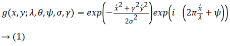
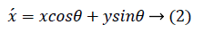
Even if the forearm blood vein images are enhanced by using adaptive histogram equalization, vein edges are might not be properly visible in most of the cases. Gabor filtering is employed to detect the edge of blood vein effectively. Image analysis using Gabor filters are often considered analogous to human visual perception as Gabor functions are proved efficient in modelling visual cortex of mammalian brains [23]. The filter can be represented in 2D spatial domain using the Equation 1 [24].
Where
and
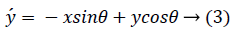
The Adaptive Histogram Equalization (AHE), which is an advancement of global histogram equalization, can be employed on such images for superior results. This advanced algorithm developed by taking account of only small regions and based on their local cumulative distribution function (CDF), performs contrast enhancement of those regions. In λ, θ, ψ, σ and γ represents the wavelength of the sinusoidal factor, orientation, phase offset, standard deviation and spatial aspect of Gabor function. Gabor filters are discussed in detail in other literatures [25-27]. This is an orientation sensitive filter used for edge detection and texture analysis. The filter is sensitive to edges in particular orientation that is given as θ value. Gabor filters with dissimilar frequencies and with orientations in different directions can be used to localize and extract blood veins from histogram equalized images. For the setup used for this research, the hand has to be placed in a specified orientation. The theta value is found and fixed as 101.247372 degrees for matching the specified orientation. This helps to segment boundaries of the major textured regions as shown in figure 6. Normalized image of the imaginary part of Gabor-filtered image is taken for further processing to achieve better results.

Figure 6: Texture enhanced image.
Blood vein extraction
For proper extraction of blood veins from the surrounding skin, selection of threshold levels is crucial. Otsu’s thresholding algorithm helps for finding suitable threshold level that efficiently extracts blood veins from the normalized Gabor filtered image. Otsu’s Thresholding performs image thresholding based on clustering [28]. By assuming the image consists of two classes of pixels, this algorithm calculates an optimal threshold value to separate the classes such that their intra-class variance is minimum and inter-class variance is maximum [29].
Otsu’s method searches for the threshold that minimizes variance within two classes as given in equation 4:
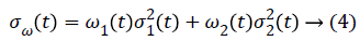
Weights ωi represents the probabilities of the two classes separated by a threshold t and σi2(t) variances of these classes. The intra-class variance is the same as maximizing inter-class variance:

Where, ωi is the class probability, and μi is the class means
From histogram, the class probability ω1 (t) is calculated as:
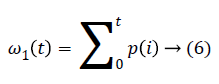
While the class mean is:
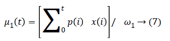
Where x(i) is the value at the center of the ith histogram. The Algorithm for the technique is explained as follows:
Find the histogram and probability of each intensity level
Setup initial 0 and 1.
Step through all possible, thresholds t=1, maximum intensity.
Update i and μi and compute σb2(t)
The desired threshold corresponds to the maximum σb2(t). Two maxima levels σb12(t) and σb22(t) are computed, and the desired threshold will be the average of two thresholds. The vein part extracted after thresholding is shown in Figure 7.

Figure 7: Resultant image after thresholding.
Shaping of blood veins
It is needed to perform morphological operations on the vein part extracted to get a more precise shape of the vein. Dilation and Erosion are mainly carried out in the post-processing for this purpose. Dilation adds pixels to the boundaries of objects in an image, while erosion removes pixels on object boundaries. Here erosion is performed, as the operation helps to remove the unwanted sections which may get extracted with the vein. The resultant image after erosion is illustrated in Figure 8. Then complement of processed image, as shown in Figure 9, is taken for inverting the black and white colours in the image.

Figure 8: Image after erosion.

Figure 9: Compliment of eroded image.
Region of interest extraction
The captured images for processing may contain portions other than forearm part which is not the region of interest. To find region of interest (ROI), the captured image is thresholded with a median pixel value around 100. This is the separating pixel value between skin and background surface. The portion of the skin where the vein is present is needed to be processed. Hence, surface part is discarded, and the region of interest is extracted. The ROI portion is inverted by fixing pixel values in the portion as 1 and all other portion as 0 is shown in Figure 10. Then the processed forearm image is multiplied with the ROI, and the resultant image is shown in figure 11.

Figure 10: Region of interest.

Figure 11: Extracted vein in the ROI.
Blood vein enhancement for real-time visibility
The remaining step is to enhance blood vein part in captured images for real-time application. The pixel value of red plane is increased to 255 in the locations corresponding to extracted veins section as shown in Figure 12.

Figure 12: Enhanced blood veins on forearm.
Results
Results of various operations performed are presented in this section. The Histogram of the original RGB image is shown in Figure 13 for a qualitative comparison with the results obtained after various operations.

Figure 13: Histogram of original image.
The qualitative similarity in the histogram of the RGB image and the histogram of the image converted to grey can be clearly observed which is shown in Figure 14. A successful intensity stretching was done with the help of contrast limited adaptive histogram equalization (CLAHE) for enhancing the intensity of the features. The efficiency of performing CLAHE twice can be clearly inferred from Figure 15, which shows the histogram of histogram equalised image.

Figure 14: Histogram of grey image.

Figure 15: Histogram of double CLAH equalised image.
The Gabor filter is used to enhance the texture features and edges for better feature extraction in the following steps. Normalized image of imaginary part of the Gabor-filtered image is used for the operations. From Figure 16, it can be clearly identified that the features in the region 0.3 to 0.5 in the normalised image are given more emphasis than other components.

Figure 16: Histogram of Gabor filtered image.
Finally, for further qualitative appreciation of the histogram of the final RGB image of enhanced extracted blood veins are shown in Figure 17. The sharp spike of red pixels represents the veins which are enhanced using red pixels with value 255. Also from the figure, it is evident that the vein features are clearly distinct from other parts of the image.

Figure 17: Histogram of RGB of enhanced extracted image.
Conclusion
Forearm blood vein identification is quite difficult for medical students during training. Apart from this doctors and nurses face tough situation to find out veins of new-born babies and senior citizens. The main reason is during unhealthy time vein visibility is very less, i.e. distinguishability between vein and skin is very less. This proposed algorithm extracted and enhanced blood vein from forearm hence providing more visibility apart from skin. The algorithm tested infrared forearm images and result shows the efficiency of this algorithm. False enhancement by this algorithm is very less and helps patients from the unwanted suffering of pain during injection in locations far from veins.
Future Works
The same algorithm can be extended for real-time vein monitoring by taking frames from real time videos and processing it. Each frame can be processed by considering them as separate images by using the same algorithm. Taking all the frames will degrade the system performance. Around 16 frames per second of the video is only required for processing, since it satisfies the law of persistence of vision. To further increase the processing speed for real-time application, it is required to use a dedicated computer having python with open CV library.
References
- Zeman HD, Lovhoiden G, Vrancken C. The Clinical Evaluation of Vein Contrast Enhancement. Proc of the 26th annual international conference of IEEE EMBS, San Francisco, CA, USA, 2004.
- Yang J, Zhang X. Feature-level fusion of fingerprint and finger-vein for personal identification. Pattern Recogn Lett 2012; 33: 623-628.
- Yang J, Shi Y. Finger-vein ROI localization and vein ridge enhancement. Pattern Recogn Lett 2012; 33: 1569-1579.
- Song W, Kim T, Kim HC, Choi JH, Kong HJ, Lee SR. A finger-vein verification system using mean curvature. Pattern Recogn Lett 2011; 32: 1541-1547.
- Zhang Z, Wang L, Zhu Q, Liu Z, Chen Y. Noise modeling and representation based classification methods for face recognition. Neurocomputing 2015; 148: 420-429.
- Kang W. Vein pattern extraction based on vectorgrams of maximal intra-neighbor difference. Pattern Recogn Lett 2012; 33: 1916-1923.
- Prasad MVNK, Kavati I, Ravindra K. Hand vein authentication system using dynamic ROI. International Symposium on Security in Computing and Communication. Springer, Berlin, Heidelberg, 2013.
- Lee JC. A novel biometric system based on palm vein image. Pattern Recogn Lett 2012; 33: 1520-1528.
- Zafar IM, Ghafoor A, Siddiqui AM. Satellite image resolution enhancement using dual-tree complex wavelet transform and nonlocal means. IEEE Geosci Remote Sens Lett 2013; 10: 451-455.
- Casaca W, Boaventura M, De Almeida MP, Nonato LG. Combining anisotropic diffusion, transport equation and texture synthesis for inpainting textured images. Pattern Recogn Lett 2014; 36: 36-45.
- Riaz MM, Ghafoor A, Sreeram V. Fuzzy C-means and principal component analysis based GPR image enhancement. In: 2013 IEEE Radar Conference (RadarCon13) 2013.
- Kono M, Ueki H, Umemura SI. Near-infrared finger vein patterns for personal identification. Appl Optics 2002; 41: 7429-7436.
- Shangqing W, Gu X. A method for hand vein recognition based on curvelet transform phase feature. Transport, Mechanical, and Electrical Engineering (TMEE), 2011 International Conference on.IEEE, 2011.
- Yihua S, Yang J. Image restoration and enhancement for finger-vein recognition. IEEE 11th International Conference on Signal Processing (ICSP), 2012.
- Gupta P, Gupta P. An accurate finger vein based verification system. Digital Signal Process 2015; 38: 43-52.
- Premalatha K, Natarajan AM. Hand Vein Pattern Recognition using Natural Image Statistics. Defence Sci J 2015; 65: 150.
- Wang J, Wang G, Li M, Du W. Hand vein recognition based on PCET. Optik - Int J Light Electron Optics 2016; 127: 7663-7669.
- Lingyu W, Leedham G. A thermal hand vein pattern verification system. International Conference on Pattern Recognition and Image Analysis. Springer, Berlin, Heidelberg, 2005.
- Qiu S, Liu Y, Zhou Y, Huang J, Nie Y. Finger-vein recognition based on dual-sliding window localization and pseudo-elliptical transformer. Expert Systems Appl 2016; 64: 618-632.
- Huang T, Yang G, Tang G. A fast two-dimensional median filtering algorithm. IEEE Transact Acoustics Speech Signal Process 1979; 27: 13-18.
- Gonzalez RC, Woods RE. Digital image processing. Nueva Jersey. 2008.
- Pizer SM, Amburn EP, Austin JD, Cromartie R, Geselowitz A, Greer T, terHaarRomeny B, Zimmerman JB, Zuiderveld K. Adaptive histogram equalization and its variations. Comput Vision Graphics Image Process 1987; 39: 355-368.
- Mar?elja S. Mathematical description of the responses of simple cortical cells. JOSA 1980; 70: 1297-1300.
- JesperJuul H. 3D surface tracking and approximation using Gabor filters. South Denmark University 2007.
- Sing LT. Image representation using 2D Gabor wavelets. IEEE Transact Pattern Anal Machine Intel 1996; 18: 959-971.
- Tan TN. Texture feature extraction via visual cortical channel modelling. Proceedings., 11th IAPR International Conference on Pattern Recognition. Vol. III. Conference C: Image, Speech and Signal Analysis, IEEE, 1992.
- David CA, Ed Jernigan M. Designing Gabor filters for optimal texture separability. Pattern Recogn 2000; 33: 1835-1849.
- Mehmet S. Survey over image thresholding techniques and quantitative performance evaluation. J Electronic Imaging 2004; 13: 146-168.
- Nobuyuki O. A threshold selection method from gray-level histograms. Automatica 1975; 11: 23-27.