ISSN: 0970-938X (Print) | 0976-1683 (Electronic)
Biomedical Research
An International Journal of Medical Sciences
Research Article - Biomedical Research (2018) Computational Life Sciences and Smarter Technological Advancement: Edition: II
A novel privacy preserving visual cryptography based scheme for telemedicine applications
Hare Ram Sah1*, G. Gunasekaran2 and Latha Parthiban3
1Faculty of Computer Science and Engineering, Sathyabama University, Chennai, India
2Department of Computer Science and Engineering, Meenakshi College of Engineering, Chennai, India
3Department of Computer Science, Pondicherry University CC, Pondicherry, India
- *Corresponding Author:
- Hare Ram Sah
Faculty of Computer Science and Engineering
Sathyabama University, Chennai, India
Accepted date: July 28, 2017
DOI: 10.4066/biomedicalresearch.29-17-519
Visit for more related articles at Biomedical ResearchWith huge quantity of patient’s medical images to be shared between specialists and clinics for improving medical diagnosis, need for privacy protection of patient details has increased. To achieve this, care must be taken that during reception, image quality must be preserved and retrieved image should be error free. In this paper, a unique concept of tiling has been used on image patterns using visual cryptography to achieve privacy preservation. The medical image is partitioned into frames using the concept of rhombus and in the deformed image tiles are formed within the different partitions, which are transmitted along with encrypted stegano-tiles. The reconstruction of original image is obtained by stacking the tiles and this error free methods performance is tested using multi scale structural similarity index and CUDA enabled GPUs.
Keywords
Visual cryptography, Tiling, Rhombus, Telemedicine
Introduction
A tiling can be periodic or non-periodic and periodic is one in which image can outline a region that tiles the plane by translation that is, by shifting the position of the region without rotating or reflecting it. Original image and the tiled image are in the same size. Hwang proposed the efficient watermark method based on Visual Cryptography (VC) and the watermark pattern can be retrieved without any information about original image [1]. Qi proposed a method to recover the secret image which is lossless [2]. Soman et al. proposed two XOR-based VC algorithms, viz., XOR-based VC for general access structure and adaptive region incrementing method [3]. Ananth et al. proposed research design that implements a barcode encoder based steganography method [4]. Linju et al. proposed the sealing algorithm where two secret images are sent at the same time by converting them to halftone representations [5]. Deepika et al. proposed a scheme to share a secret among ‘n’ participants, i.e. an ‘n’ out of ‘n’ secret sharing scheme, based on a new number system called Permutation Ordered Binary (POB) number system and Chinese Remainder Theorem (CRT) [6]. Niimi et al. proposed a method for preserving privacy in electronic health record [7]. Parallel cryptosystem is introduced, using which all subtiles are transmitted at the same time and in, an algorithm to for encryption of secret images into meaningful images is proposed [8,9].
Proposed Methodology
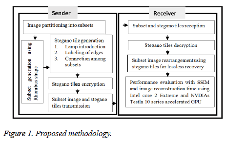
The proposed privacy preserving method is achieved by partitioning the medical image by tiling, applying stegano algorithm followed by simple encryption which is decrypted in receiving end as shown in Figure 1.
Partitioning the medical image by tiling
Tiling is a way of arranging identical plane shapes, so that they completely cover an area without overlapping. The rhombus based partition is placed over the secret image. The secret image is divided into six subsets based on the angles used in Figure 2 and partitioned as shown in Figure 3.
Applying stegano algorithm to generate stegano tiles
The steps for stegano algorithm are given by:
• Introduce lamps in secret figure.
• Label the edges of the subsets as Hik, i=1 to L (number of subsets) and k=1 to number of edges in ith tile.
• The connecting among subsets specified in the stegano tile.
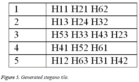
Lamps are introduced to rhombus shape and the generated five lamps and labelling process are shown in Figure 4. The generated stegano tile is shown in Figure 5.
Encryption of stegano tile
The encryption is based on the attribute of database and if the data type of attribute is text, then it is encrypted into numerical value and if the data type of attribute is numeric, then it is encrypted into text as in Table 1.
| Text attribute | Key | Text attribute | Key | Text attribute | Key | Text attribute | Key | Text/Numeric attribute | Key | Numeric attribute | Key |
|---|---|---|---|---|---|---|---|---|---|---|---|
| A | 1 | G | 7 | M | 13 | S | 19 | Y | 25 | 4 | E |
| B | 2 | H | 8 | N | 14 | T | 20 | Z | 26 | 5 | F |
| C | 3 | I | 9 | O | 15 | U | 21 | 0 | A | 6 | G |
| D | 4 | J | 10 | P | 16 | V | 22 | 1 | B | 7 | H |
| E | 5 | K | 11 | Q | 17 | W | 23 | 2 | C | 8 | I |
| F | 6 | L | 12 | R | 18 | X | 24 | 3 | D | 9 | J |
Table 1. Encryption of text and numeric attribute.
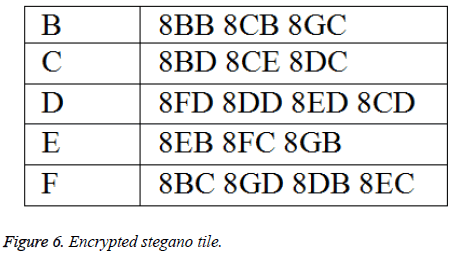
The attribute based encryption technique is applied to the Figure 6 and the encrypted stegano tile is shown in Figure 6. This encrypted stegano tile along with labelled tiles are received in the receiving end which is decrypted (same as Figure 5) and help in the reconstruction of the original image.
Results and Discussions
The proposed algorithm is tested on the CT image (Figure 7) which is partitioned based on the procedure in Figure 3.
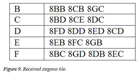
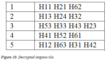
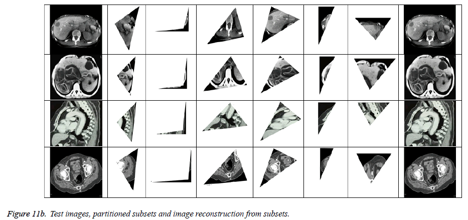
The generated tiles are labelled based on rhombus shape as shown in Figure 8 which is then encrypted (Figure 6) and transmitted. The received stegano tile in encrypted form is shown in Figure 9 and decrypted stegano tile is shown in Figure 10. In reconstruction process (Figures 11a and 11b), the labelled tiles are joined in step by step manner from decrypted stegano tile (Figure 6) to obtain the secret image.
The proposed algorithm was tested on the 4 CT medical images (obtained from www.aylward.org/notes/open-accessmedical-image-repositories) and the reconstructed images are compared with original images in terms of Multi-scale Structural Similarity Index (SSIM). Table 2 shows the SSIM obtained for the 4 test images.
| Test Image | SSIM |
|---|---|
| 1 | 0.9878 |
| 2 | 0.9912 |
| 3 | 0.9868 |
| 4 | 0.9976 |
Table 2. SSIM results for the test images.
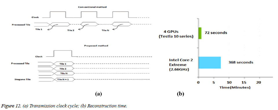
The proposed method is not dependent on the order of arrival of tiles like the conventional method as in Figure 12 (a). NVIDIA’s Tesla GPUs reconstruction time performance of the partitioned subsets using encrypted stegano tile was found to be only 72 seconds against 368 seconds taken by Intel’s core 2 Extreme as shown in Figure 12 (b).
Conclusions
In this paper as reconstruction of original medical image is done by stacking the stegano tiles, it is an enhanced privacy preserving lossless technique for e-health applications. The method is easy to implement and does not need complicated cryptographic computations. The received images are found to have multi scale structural similarity nearing 1. The reconstruction time was also found to be very less when CUDA enabled GPUs were used. Future work is to concentrate on other efficient tiling shapes and use other Testla processors for faster reconstruction time.
Acknowledgement
The authors would like to thank Pondicherry University for allowing access to CUDA enabled GPU computing facility for the research work.
References
- Ren-Junn H. A digital image copyright protection scheme based on visual cryptography. Tamkang J Sci Eng 2000; 3: 97-106.
- Xin Q. Lossless recovery of multiple decryption capability and progressive visual secret sharing. Int J Grid Dist Comp 2016; 9: 51-60.
- Nidhin S, Smruthy B. XOR-based visual cryptography. Int J Cybernet Inform 2016; 5: 253-264.
- Vijay AS, Sudhakar P. Performance analysis of a combined cryptographic and steganographic method over thermal images using barcode encoder. Indian J Sci Technol 2016; 9: 1-5.
- Linju PS, Sophiya M. An efficient interception mechanism against cheating in visual cryptography with non-pixel expansion of images. Int J Sci Technol Res 2016; 5: 102-106.
- Deepika MP, Sreekumar A. A novel secret sharing scheme using POB number system and CRT. Int J Appl Eng Res 2016; 11: 2049-2054.
- Niimi, Yukari O, Katsumasa. Examination of an electronic patient record display method to protect patient information privacy. CIN: Comp Inform Nurs 2017; 35: 100-108.
- Hong-Mei Y, Ye L, Tao L, Ting H, Li-Hua G. A new parallel image cryptosystem based on 5D hyper-chaotic system. Signal Processing: Image Commun 2017; 52: 87-96.
- Kanso A, Ghebleh M. An algorithm for encryption of secret images into meaningful images. Optics Lasers Eng 2017; 90: 196-208.
