Biomedical Research
International Journal of Pediatrics
Down syndrome: Cryptic paternal translocation (9;21).
1Department of Research on Children with Special Needs, National Research Center.
2Department of Human Cytogenetics, National Research Center, Cairo, Egypt
- *Corresponding Author:
- Nagwa Abdel Meguid
Section of Human Genetics
National Research Center, Tahrir street
Dokki, Cairo, Egypt
Accepted Date: February 01 2012
Down syndrome is the most frequent autosome aneuploidy in live newborns, caused by trisomy of all or the critical region of chromosome 21. We described a 20 month old boy with Down syndrome phenotype. Full clinical examination was done. Cytogenetic studies for the proband and his parents using GTG- banding technique and Fluorescence Insitu Hybridization technique using LSI 21 and chromosome 9q subtelomere probe were used according to the manufacturer’s instructions. The proband;s karyotype is: 46,XY,der(9)t(9;21)(p24;q22.2) resulted from paternal balanced cryptic translocation. Karyotype of the father is 46,XY, t(9;21)(p24;q22.2) , while mother showed normal Karyotype. FISH analysis using LSI 21 probe and 9q subtelomere probe for the proband’s and father demonstrated the presence of three signals, and two signals respectively. This is the first report describing a paternal cryptic balanced translocation involving chromosome 9 with the generation of aneuploidy contributing to abnormal chromosomal segregation.
Keywords
Down syndrome, cryptic translocation, FISH
Introduction
Down syndrome DS the most frequent form of mental retardation caused by a chromosomal aberration is characterized by well-defined and distinctive phenotypic features and caused by trisomy of all or a critical portion of chromosome 21 with a frequency of 1 in 650 to 1,000 live births in the population.
It has long been recognized that the risk of having a child with trisomy 21 increases with maternal age; the risk of having a liveborn with DS at maternal age 30 is 1 in 1,000 and at maternal age 40 is 9 in 1,000.
Reviewed studies [1,2,3,4,5] reported errors in meiosis that lead to trisomy 21 were overwhelmingly of maternal origin; only 5% occur during spermatogenesis, while most errors occur in meiosis I which account for 76 to 80% and maternal meiosis II errors constitute 20 to 24%. In families in which there is paternal nondisjunction, most of the errors occur in meiosis II, while in 5% of trisomic individuals the supernumerary chromosome 21 appears to result from an error in mitosis. These data were also supported in many studies as approximately 91% of DS cases arise from maternal nondisjunction (ND) and only 9% from paternal aneuploidies.[6] All de novo t (14; 21) trisomies have originated in maternal germ cells, [7] the mean maternal age was 29.2 years. In de novo t (21; 21) DS the situation is different, [8,9,10] in most cases the t (21; 21) is an isochromosome (dup 21q) rather than the result of a Robertsonian translocation caused by a fusion between 2 heterologous chromatids. About half were of paternal and half of maternal origin. Less frequently t (21; 21) true Robertsonian trisomy 21 cases, the extra chromosome 21 was maternal.
Here we report on a family who had a child with DS due to paternal ND in Meiosis II, FISH analysis using the LSI 21 probe (Vysis ) spectrum orange (21q22.13-q22.2) showed that the father had an extra positive signal on chromosome 9. We consider being a cryptic translocation, which seems to be related to the paternal ND that gave rise to two sibs with DS.
Clinical Examination
A male patient aged one year and 8ms. at time of examination was referred to the clinic of Children with Special Needs, National Research Center having DS. The parents were healthy, nonconsanguineous, under 35 years of age when the DS child was born, and had another DS daughter. History of a previous child diagnosed as DS who died at the age of 5m and a paternal female cousin with DS.
The proband was delivered by C.S. (elective), ± full term after a pregnancy with threatened abortion during the second month of gestation. Physical examination showed a classical DS phenotype with weight 25th centile, length 10th centile, head circumference 3rd centile, mild brachycephaly, flat nasal bridge, upward slanting palpebral fissures, epicanthal folds, asymmetric dysplastic ears with folded right helix, high arched palate, micrognathia, loose skin on neck nape, small genitalia, brachydactyly, single transverse palmar crease and gap between first and second toes. Echo Cardiography demonstrated the presence of VSD.
Cytogenetic studies and Results
Chromosomal analysis for the family was done using Gbanding technique according to the described methods, a total of 25 metaphases were analyzed for each case [11,12]. Structural or numerical anomalies were recorded and karyotyped according to the ISCN (2009). [13] Cytogenetic analysis of the proband revealed 46, XY, der(9)t(9;21)(p24;q22.2) , Fig. (1). Cytogenetic analysis for the sister was not available, mother showed normal Karyotype 46, XX and father showed 46, XY, t(9;21)(p24;q22.2) respectively.
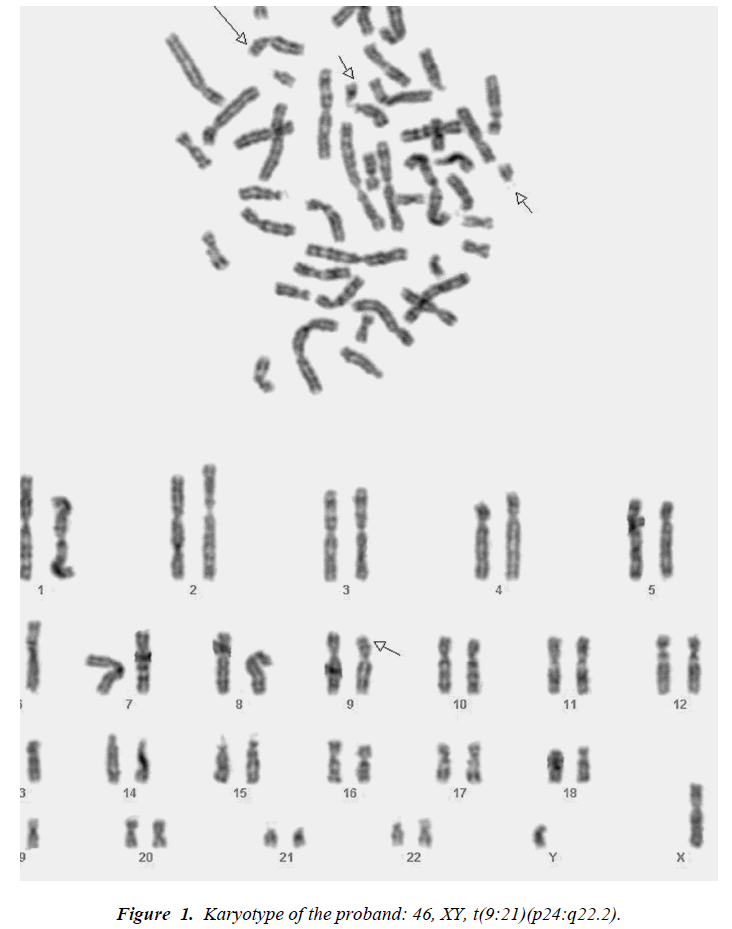
Figure 1: Karyotype of the proband: 46, XY, t(9:21)(p24:q22.2).
Fluorescent in situ hybridization (FISH) using LSI 21 probe (Vysis ) spectrum orange (21q22.13-q22.2) to identify the critical region of Down syndrome and Tel. 9q probe (Vysis) spectrum orange to identify the long arm of chromosome 9 was applied. [14] Slides prepared from 3:1 methanol: acetic acid fixed cells, pretreated with 2xSSC at 37° C for 30 min., dehydrated for 2 min. in 70%, 90% and 100% ethanol respectively. Denaturation of the slides in 70% formamide at 73 ± 1° C for 5 min. were done followed by dehydration in cold ethanol 70%, 90% and 100%. Denaturation of 10 μl of the probe in a water bath at 74 ± 1 °C for 5 min. was done, added to slides, covered and sealed with rubber cement and incubated at 37°C overnight. Slides were FISH using LSI 21 probe (Vysis ) (spectrum orange) of the proband demonstrating the presence of 3 signals as indicated by the arrows Fig. (2), two signals represent normal signals on chromosome 21q22.2 while the third one represent the translocated signal to the short arm of chromosome 9 (9p24). FISH of the mother using LSI 21 probe (Vysis) (spectrum orange) (arrow heads) and Tel. 9q probe (Vysis) (spectrum orange) (arrows) revealing normal signals. Fig. (3). FISH of the father using LSI 21 probe (Vysis ) spectrum orange (short arrow ) showed two signals, one signal on 21q22.2 and a translocated signal to the terminal end of the short arm of chromosome 9 (9p24) Fig. (4).

Figure 2: FISH using LSI 21 probe (Vysis ) (spectrum orange) of the proband demonstrating the presence of 3 signals.
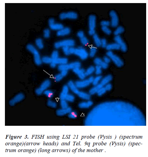
Figure 3: FISH using LSI 21 probe (Vysis ) (spectrum orange)(arrow heads) and Tel. 9q probe (Vysis) (spectrum orange) (long arrows) of the mother .
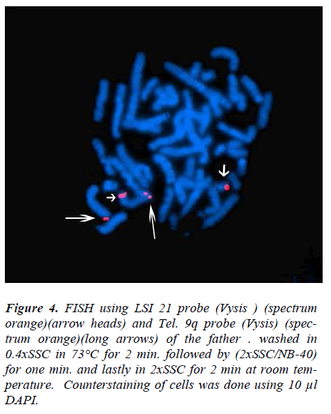
Figure 4: FISH using LSI 21 probe (Vysis ) (spectrum orange)(arrow heads) and Tel. 9q probe (Vysis) (spectrum orange)(long arrows) of the father . washed in 0.4xSSC in 73°C for 2 min. followed by (2xSSC/NB-40) for one min. and lastly in 2xSSC for 2 min at room temperature. Counterstaining of cells was done using 10 μl DAPI.
Discussion
To date, advanced maternal age is the only factor confirmed to be associated with chromosome 21 ND, while ND of paternal origin shows no association with advanced age. A relatively small region may play a major role in DS phenotypes, and proposed the concept of a DSCR [15] which was defined with a proximal boundary between markers D21S17 (35 892 kb) and D21S55 (38 012 kb), and a distal boundary at MX1 (41 720 kb) [16,17].
We present a case with DS phenotype where cytogenetic examination revealed 46, XY, t(9:21)(p24:q22.2). FISH examination demonstrated the presence of a translocated signal to the short arm of chromosome No.9. Parental examination has revealed normal 46, XX for the mother and 46, XY for the father while FISH analysis revealed a translocated signal to 9p proofing that segregation is an apparently balanced cryptic translocation from the father.
Partial duplication of the DSCR (D21S55, sub band 21q22.2) of maternal origin in the patient was documented; [18] the trisomy was due to segregation of an apparently balanced cryptic translocation from the mother. The patient's karyotype was 46,XX,- 12,tder(12)t(12;21)(p13.1;q22.2)maternal. In our study the father showed a balanced translocation between 21q22.2 and 9p24 as evidenced from the normal size of the two chromosomes 21, however the possibility of other chromosome translocations cannot be excluded. The cryptic translocation found on the paternal der (9) chromosome could have led to ND of chromosome 21 in sperm, giving rise to a DS child. Gair et al. [2005] suggested that a cryptic translocation on chromosome 21 may have increased the frequency of ND of this chromosome in a family with recurrent trisomy 21 in three generations [19].
Two cases were reported with partial trisomy 21 having the clinical features distinct for DS, each patient had a similarly sized extra chromosome 21 , each of their extra chromosomes contained a distal part of chromosome 3p or 14q at the telomeric region of chromosome 21q ( from maternal origin) [20]. In another study , they reported many individuals displaying subtle translocations and internal rearrangements causing duplications and deletions of HSA21 region, for some patients additional copies of several non-contiguous regions were detected indicating that complex rearrangements involving multiple events had occurred. [21] The present study represents a rare balanced translocation in a proband in a DS family where the origin is from a cryptic paternal translocation as most reported familial non- acrocentric translocation cases were of maternal origin and also highly evaluate the use of different FISH techniques for proper diagnosis and genetic counseling. It is important to establish the frequency of cryptic translocations, which may be an important etiologic factor in the generation of the most frequent chromosomal syndromes in future generations.
References
- Antonarakis, S. E.(1993): Human chromosome 21: genome mapping and exploration circa 1993. Trends Genet. 9: 142-148.
- Antonarakis, S. E., Lewis, J. G., Adelsberger, P. A., Petersen, M. B., Schinzel, A. A., Binkert, F., Schmid, W., Pangalos, C., Raoul, O., Chakravarti, A., Hafez, M., Cohen, M. M., Roulston, D., Schwartz, S., Mikkelsen, M., Tranebjaerg, L., Greenberg, F., Hoar, D. I., Rudd, N. L., Warren, A. C., Metaxotou, C., Bartsocas, C., (1991): Down Syndrome Collaborative Group Parental origin of the extra chromosome in trisomy 21 using DNA polymorphism analysis. New Eng. J. Med. 324: 872-876.
- Antonarakis, S. E., Petersen, M. B., McInnis, M. G., Adelsberger, P. A., Schinzel, A. A., Binkert, F., Pangalos, C., Raoul, O., Slaugenhaupt, S. A., Hafez, M., Cohen, M. M., Roulson, D., Schwartz, S., Mikkelsen, M., Tranebjaerg, L., Greenberg, F., Hoar, D. I., Rudd, N. L., Warren, A. C., Metaxotou, C., Bartsocas, C., Chakravarti, A.(1992): The meiotic stage of nondisjunction in trisomy 21: determination using DNA polymorphisms. Am. J. Hum. Genet. 50: 544-550.
- Sherman, S. L., Freeman, S. B., Grantham, M., Peters, J., Jacobs, P. A., Kurnit, D. M., Uchida, I., Petersen, M. B., Mikkelsen, M., Hassold, T. J. (1992): Nondisjunction of trisomy 21: comparison of centromere maps resulting from maternal meiosis I and II nondisjunction. Am. J. Hum. Genet. 51 (suppl.): A24.
- Sherman, S. L., Takaesu, N., Freeman, S. B., Grantham, M., Phillips, C., Blackston, R. D., Jacobs, P. A., Cockwell, A. E. (1991): Association between reduced recombination and nondisjunction. Am. J. Hum. Genet. 49: 608-620.
- Zaragoza MV, Jacobs PA, James RS, Rogan P, Sherman S, Hassold T. (1994): Nondisjunction of human acrocentric chromosomes: studies of 432 trisomic fetuses and liveborns. Hum Genet. Oct;94(4):411-7.
- Petersen, M. B., Adelsberger, P. A., Schinzel, A. A., Binkert, F., Hinkel, G. K., Antonarakis, S. E. (1991): Down syndrome due to de novo Robertsonian translocation t14;21: DNA polymorphism analysis suggests that the origin of the extra 21q is maternal. Am. J. Hum. Genet. 49: 529-536.
- Shaffer, L. G., Jackson-Cook, C. K., Stasiowski, B. A., Spence, J. E., Brown, J. A. (1992): Parental origin determination in 30 de novo Robertsonian translocations. Am. J. Med. Genet. 43: 957-963.
- Grasso, M., Giovannucci, M. L., Pierluigi, M., Tavellini, F., Perroni, L., Dagna-Bricarelli, F. (1989): Isochromosome, not translocation in trisomy 21q21q. Hum. Genet. 84: 63-65.
- Antonarakis, S. E., Adelsberger, P. A., Petersen, M. B., Binkert, F., Schinzel, A. A. (1990): Analysis of DNA polymorphism suggests that most de novo dup(21q) chromosomes in patients with Down syndrome are isochromosomes and not translocations. Am. J. Hum. Genet. 47: 968-972.
- Seabright M (1971): A rapid banding technique for human chromosomes. Lancet, 30; 2(7731): 971-2.
- Verma R.S and Babu A (1995): Human chromosomes: Principal and Techniques. 2nd ed. New York, San Francisco, Mc Graw-Hill Inc.
- ISCN (2009): An International System of Human Cytogenetic Nomenclature, Shaffer L. (Ed.), Karger, Basel, Krager.
- Pinkel D, Gray J, Trask B, Van den Engh G, Fuscoe J and Van Dekken H.(1986): Cytogenetic analysis by in situ hybridization with fluorescently labeled nucleic acid probes. Cold Spring Harbor Symp Quant Bio. 51:151-157.
- Sinet PM, Theophile D, Rahmani Z, et al. (1994): Mapping of the Down syndrome phenotype on chromosome 21 at the molecular level. Biomed Pharmacother. 48:247.
- Delabar, J. M., Theophile, D., Rahmani, Z., Chettouh, Z., Blouin, J. L., Prieur, M., Noel, B., Sinet, P. M. (1993): Molecular mapping of twenty-four features of Down syndrome on chromosome 21. Europ. J. Hum. Genet. 1: 114-124.
- Korenberg, J. R., Chen, X.-N., Schipper, R., Sun, Z., Gonsky, R., Gerwehr, S., Carpenter, N., Daumer, C., Dignan, P., Disteche, C., Graham, J. M., Jr., Hugdins, L., McGillivray, B., Miyazaki, K., Ogasawara, N., Park, J. P., Pagon, R., Pueschel, S., Sack, G., Say, B., Schuffenhauer, S., Soukup, S., Yamanaka, T.(1994): Down syndrome phenotypes: the consequences of chromosomal imbalance. Proc. Nat. Acad. Sci. 91: 4997-5001.
- Scott JA, Wenger SL, Steele MW, Chakravarti A. (1995): Down syndrome consequent to a cryptic maternal 12p;21q chromosome translocation. Am J Med Genet. Mar 13;56(1):67-71.
- Gair JL, Arbour L, Rupps R, Jiang R, Bruye`re H, Robinson WP. (2005): Recurrent trisomy 21: Four cases in three generations. Clin Genet 68:430–435.
- Kondo Y, Mizuno S, Ohara K, Nakamura T, Yamada K, Yamamori S, Hayakawa C, Ishii T, Yamada Y, Wakamatsu N.(2006): Two cases of partial trisomy 21 (pter-q22.1) without the major features of Down syndrome. Am J Med Genet A. Feb 1;140(3):227-32.
- Korbel JO, Tirosh-Wagner T, Urban AE, Chen XN, Kasowski M, Dai L, Grubert F, Erdman C, Gao MC, Lange K, Sobel EM, Barlow GM, Aylsworth AS, Carpenter NJ, Clark RD, Cohen MY, Doran E, Falik- Zaccai T, Lewin SO, Lott IT, McGillivray BC, Moeschler JB, Pettenati MJ, Pueschel SM, Rao KW, Shaffer LG, Shohat M, Van Riper AJ, Warburton D, Weissman S, Gerstein MB, Snyder M, Korenberg JR. (2009): The genetic architecture of Down syndrome phenotypes revealed by high-resolution analysis of human segmental trisomies. Proc Natl Acad Sci USA à106:12031–12036.