ISSN: 0970-938X (Print) | 0976-1683 (Electronic)
Biomedical Research
An International Journal of Medical Sciences
Case Report - Biomedical Research (2017) Volume 28, Issue 8
Sigmoid colon perforation due to a jujube pit
Department of Gastrointestinal Surgery, Affiliated Hospital of Putian University, PR China
- *Corresponding Author:
- Jianxin Chen
Department of Gastrointestinal Surgery
Affiliated Hospital of Putian University, PR China
Accepted date: January 03, 2017
Jujube is a common fruit in Chinese diet, jujube pit is sharp but has rarely been reported to cause digestive tract perforation. Here we presented a rare case of sigmoid colon perforation by a jujube pit in an 88 year-old toothless woman. The evidence of peritoneal irritation was acquired by physical examination and CT scan. Then perforation peritonitis was diagnosed, and emergency operation was performed accordingly. The patient recovered smoothly and was discharged after 10 days of hospital stay.
Keywords
Peritonitis, Perforation, Foreign body, Jujube
Introduction
Most foreign bodies pass through the digestive tract without serious incidents. Only a few sharp foreign bodies can perforate the bowel such as fish bones, toothpicks, olive seeds, glass shard and pear seeds [1]. Less than 1% of ingested foreign bodies will perforate the bowel, and the greatest risk is with large and sharp objects. Here we present a case of sigmoid colon perforation due to a jujube pit, which has rare been reported before.
Case Report
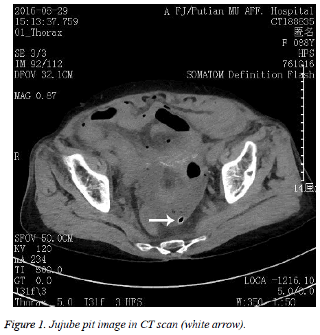
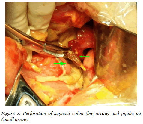
An 88 year-old woman whose teeth had been all lost visited our hospital with complaint of abdominal pain. The process of disease lasted thirty hours, and her symptom became serious gradually. Physical examination revealed the evidence of peritoneal irritation: white blood cell count (WBC) was 16.72 × 10⁹/L (normal range 3.5-10.5 × 10⁹/L), neutrophilia was 85.8%, C-reactive protein level was elevated to 200 mg/L (normal<10 mg/L), and procalcitonin level was elevated to 100 ng/ml (normal<0.5 ng/ml). An abdominal CT scan showed a sharp foreign body in low left quarter of abdominal cavity (Figure 1). A diagnosis of perforation peritonitis due to foreign body was made and an emergency exploratory operation was performed. A perforation was found in sigmoid colon by a sharp jujube pit (Figure 2). After cleaning the abdominal cavity, local colon resection, colostomy and drainage of the abscess were performed. Post-operative pathology revealed part colon with suppurated inflammation. The patient was discharged after ten days of hospital stay.
Discussion
The accidental ingestion of foreign body is mainly among children, adolescents, the elderly and patients with dental problems. The most frequent sites of perforation by foreign body are the ileocecal and rectosigmoid regions due to narrow bowel cavity and angled digestive tract [1,2]. Many fruits such as apricots, persimmons and plums have previously been described in the literature as a cause of intestinal obstruction, either through direct mechanical obstruction by the pit or through formation of a phytobezoar [3-5].
The jujube is a fruit of the jujube tree which originally grows in China. The jujube pit is fusiformis body with two sharp sides. This fruit are often seen mixed with rice in Chinese diet. The patient had lost all teeth and unable to chew carefully, so she swallowed the pit by accident during meal. Then, the jujube pit caused a bowel perforation in sigmoid colon. To our knowledge, this is the first report of jujube pit perforation of sigmoid colon at Affiliated Hospital of Putian University. When a foreign body is suspected, endoscopy has the advantage of noninvasive examination and removal. Examination with flexible endoscope for upper digestive tract is considered an accurate diagnostic measure [6]. However, the use of endoscopy is limited when the disease is associated with perforation peritonitis. Acute perforation is usually clinically remarkable, and the history is short as patients present with acute peritonitis. If the foreign body is radiopaque, X-rays can be used to locate the object. Plain radiography is helpful in locating metallic foreign body, but it is not useful in locating other foreign bodies such as chicken or fish bones. CT scan is helpful in recognizing intestinal perforation caused by jujube pit, which usually presents as a fusiformis foreign body with external high density and internal low density. Furthermore, CT scan allows the differential diagnosis of other causes of abdominal pain.
Typical treatments for foreign-body perforation are drainage of the abscess and use of antibiotics with or without bowel resection [7]. The lack of intra-abdominal contamination and the limited range of the lesion allowed performing of simple suture [8]. When perforation affects colon, colostomy is generally recommended because these cases usually have abdominal cavity contamination and most of this cases have diffuse peritonitis [9]. In this case, the perforation had caused diffuse peritonitis. Local resection, colostomy and drainage of the abscess were suitable to perform in the patient. In summary, ingestion of jujube pit may cause serious perforation of the bowel. CT scan is a helpful examination for the diagnosis of this disease. An operation should be performed immediately.
References
- Goh BK, Chow PK, Quah HM, Ong HS, Eu KW, Ooi LL, Wong WK. Perforation of the gastrointestinal tract secondary to ingestion of foreign bodies. World J Surg 2006; 30: 372-377.
- Kornprat P, Langner C, Mohadjer D, Mischinger JH. Chicken-bone perforation of a sigmoid colon diverticulum into the right groin and subsequent phlegmonous inflammation of the abdominal wall. Wien Klin Wochenschr 2009; 121: 220-222.
- Atila K, Güler S, Bora S, Gülay H. An unusual cause of small bowel perforation: apricot pit. Ulus Travma Acil Cerrahi Derg. 2011; 17: 286-288.
- Delia CW. Phytobezoars (Diospyrobezoars). A clinicopathologic correlation and review of six cases. Arch Surg 1961; 82: 579-583.
- Zelman S. The case of the perilous prune pit. N Engl J Med 1961; 265: 1202-1204.
- Brady PG, Johnson WF. Removal of foreign bodies: the flexible fiberoptic endoscope. South Med J 1977; 70: 702-704.
- Yamamoto M, Yamamoto K, Sasaki T, Fukumori D, Yamamoto F, Igimi H, Yamamoto H, Yamashita Y. Successfully Treated Intra-Abdominal Abscess Caused by Fish Bone With Perforation of Ascending Colon: A Case Report. Int Surg 2015; 100: 428-430.
- Pinero Madrona A, Femández Hernández JA, Carrasco Prats M, Riquelme J, Parrila Paricio P. Intestinal perforation by foreign bodies. Eur J Surg 2000; 166: 307-309.
- Rodríguez-Hermosa JI, Codina-Cazador A, Sirvent JM, Martín A, Gironès J, Garsot E. Surgically treated perforations of the gastrointestinal tract caused by ingested foreign bodies. Colorectal Dis 2008; 10: 701-707.

