ISSN: 0970-938X (Print) | 0976-1683 (Electronic)
Biomedical Research
An International Journal of Medical Sciences
Research Article - Biomedical Research (2017) Volume 28, Issue 10
Identification and analysis of toll like receptor 4 (TLR4) level changes in vascular dementia patients related type 2 diabetes mellitus
Fan Yaochun1,3#, Ou Xia2#, Wang Wenrui3, Tian Xiaoling3, Yan Shaohong1,3, Hu Nan1, Zhang Xiaoyan1 and Xing Wanjin1*
1College of Life Sciences of Inner Mongolia University, No.235 of West Road, Saihan District, Hohhot, Inner Mongolia, PR China
2Basic Medical College of Kunming Medical University, 1168 of Chunrong West Road, Yuhua Street, Chenggong New Town, Kunming, Yunnan, PR China
3Inner Mongolia Autonomous Region Comprehensive Center for Disease Control and Prevention; Address: No. 50 of Erdos Street, Yuquan District, Hohhot, Inner Mongolia, PR China
#These authors contributed equally to this work
- *Corresponding Author:
- Xing Wanjin
College of Life Sciences of Inner Mongolia University
Inner Mongolia, PR China
Accepted date: March 4, 2017
Objective: Since the Vascular Dementia (VD) is one of serious complication of Type 2 Diabetes Mellitus (T2DM), we study the molecular mechanism of Toll Like Receptor 4 (TLR4) in autoimmunity respond on VD patients.
Methods: The clinical symptoms of VD related T2DM patients are investigated and VD risk factors are analysed. We exam the blood samples by immunofluorescence assay. After collecting the patients’ Cerebrospinal Fluid (CSF), it tests the level of TLR4 in CSF and blood at gene and protein degrees, respectively. Flow cytometry is used to verify the higher level of TLR4 play an important role in the autoimmune response of VD patients.
Results: The average incidence of T2DM is 1.36%, and VD incidence is 0.33% in 2016. The VD patients are concentrated in old and middle-age women. The risk factors of VD are including rural residence, inadequate health-management, >7 years of T2DM-history and mental pressure. TLR4 protein were at higher presence in VD groups than the normal T2DM group and control group, which consistent with the results of TLR4 mRNA expression in CSF. Additional, their CD4+/CD8+ ratio is increased significantly, associated with TLR4 increasing expression.
Conclusion: VD is directly related with TLR4 increasing expression, which is a threat to the health of the T2DM patients.
Keywords
Type 2 diabetes mellitus, Toll like receptor 4, Vascular dementia, Expression
Introduction
Type 2 Diabetes Mellitus (T2DM) is a long term metabolic disorder that is characterized by high blood sugar, insulin resistance, and relative lack of insulin [1-3]. The normal T2DM patients have some symptoms, including increased thirst, frequent urination, and unexplained weight loss. Although the disease progressed slowly, these long-term complications from high blood sugar include heart disease, strokes, diabetic retinopathy, resulting blindness, kidney failure, and poor blood flow in the limbs which may lead to amputations [4]. One of the most important complication is Vascular Dementia (VD), also known as multi-infarct dementia (MID) and Vascular Cognitive Impairment (VCI), is dementia caused by problems in the supply of blood to the brain, typically a series of neuronal inflammation, leading to worsening cognitive decline when the progressive development of the disease [5,6].
Vascular dementia can sometimes be triggered by cerebral amyloid angiopathy, which involves accumulation of beta amyloid plaques in the walls of the cerebral arteries, leading to breakdown and rupture of the vessels. Since amyloid plaques are a characteristic feature of Alzheimer's disease, vascular dementia may occur as a consequence [7]. However, cerebral amyloid angiopathy can appear in people with no prior dementia condition, e.g.T2DM patients.
The VD related T2DM refers to a syndrome consisting of a complex interaction of cerebrovascular disease and risk factors that lead to changes in the brain structures due to inflammation and lesions, and resulting changes in cognition [6]. The temporal relationship between a self-inflammatory response and cognitive deficits is needed to make more research in this study, which focused on TLR4.
The protein with TLR4 is a member of the Toll-Like Receptor (TLR) family, which plays a fundamental role in pathogen recognition and activation of innate immunity [8]. TLRs are highly conserved from drosophila to humans and share structural and functional similarities [9]. They recognize Pathogen-Associated Molecular Patterns (PAMPs) that are expressed on infectious agents, and mediate the production of cytokines necessary for the development of effective immunity [8,9]. At the same time, the various TLRs exhibit different patterns of expression. This receptor is most abundantly expressed in placenta, and in myelomonocytic sub-population of the leukocytes.
This research is to confirm the function of TLR4 in the progressive development of VD related T2DM about cognitive dysfunction. On the other hand, T2DM is also associated with the development of cognitive dysfunction in vascular dementia (risk factors such as age, duration of diabetes, genetic factors, blood pressure, etc.). In the TLR4-mediated self-inflammatory response, T2DM is one of the main risk factors in VD cognitive impairment.
Material and Methods
Patients and diagnosis
It collected 177 T2DM patients from the Peking Union Hospital from January to December, 2016. They were received treatment and diagnosis from the Department of Endocrinology. The specific details of the diagnosis criteria were according to the Guideline of World Health Organization (WHO) [10]. All initiatives of definition, diagnosis and classification of diabetes mellitus and their complications were list in Table 1.
| Condition | 2 h glucose | Fasting glucose | HbA1c | |
|---|---|---|---|---|
| Unit | mmol/l (mg/dl) | mmol/l (mg/dl) | mmol/mol | DCCT % |
| Normal | <7.8 (<140) | <6.1 (<110) | <42 | <6.0 |
| Impaired fasting glycaemia | <7.8 (<140) | ≥ 6.1 (≥ 110) and <7.0 (<126) | 42-46 | 6.0-6.4 |
| Impaired glucose tolerance | ≥ 7.8 (≥ 140) | <7.0 (<126) | 42-46 | 6.0-6.4 |
| Diabetes mellitus | ≥ 11.1 (≥ 200) | ≥ 7.0 (≥ 126) | ≥ 48 | ≥ 6.5 |
Table 1. The WHO criteria of diagnosis and classification of diabetes mellitus.
Vascular Dementia (VD), the specific diagnostic criteria were used the diagnostic and statistical manual of mental disorders to diagnose VD. And all 43 VD patients were exam with the Hachinski Ischemic Score [11]. If their scores were above 7, they were allocated to VD group, otherwise Non VD patients group. It investigated all patients’ epidemiological data and taken pathogenic spectrum testing if necessary.
Patients’ symptoms
The patients’ general symptoms were distributed as following: 159 cases were with frequent urination, 144 cases with increased thirst, 119 cases with increased hunger, and 121 cases with weight loss (up to 20% losing at most).
Other indirect complications were as following: 97 cases were with blurred vision, 99 cases with itchiness, 81 cases with peripheral neuropathy, and 38 cases with recurrent vaginal infections. Especially, it found 17 cases with hyperosmolar hyperglycemic state, whose condition of most high blood sugar, were associated with a decreased level of consciousness and low blood pressure.
Signs and symptoms of VD
By the early manifestations of VD, they were with normal condition of cognitive, motor, behavioral and affective. But all changes typically occurred over a period of 7 to 10 years. Signs were mainly included cognitive decline (86.52%) and memory impairment (78.95%), which interferes with their daily living. A total of 33 VD patients with focal neurologic signs, by evidence of features consistent with cerebrovascular disease on brain imaging (CT and MRI). When they have patchy deficits in terms of cognitive testing, they tended to have better free recall and fewer recall intrusions when compared with patients with Alzheimer's disease. In the more severe patients, only one case presented dysarthrias and aphasias.
Control group
The 160 patients were suffered from non-T2DM diseases over the same period in the same hospital. All these control cases were randomly selected, without any other restrictions. These participants provided written informed consent. This study was according to the declaration of Helsinki and world health organization guidelines to implement. It was under the supervision of the Ethics Committee of the Peking Union Medical College (No.PUCCMMU201101009149). The patients’ data were recorded, including symptoms, history of prior disease and physical examination. Even discharged from hospital, the physicians performed a monthly telephone following-up.
Detection of blood
After fully informed, we recruited 177 T2DM patients and 160 control patients for this research experiment. By patients’ last visiting hospital, it collected 2-3 ml fasting venous blood, then separated the serum by centrifugal for each experimental group and the control group. Saved their serum in -20°C and detected their insulin related index for further. The Immunofluorescence Assay (IFA) kits were purchased from the Shanghai enzyme of the Biological Technology Co., Ltd (Shanghai, China) (Cat.No.ml022831). According to its manual, all assays were implemented.
Lumbar puncture
Lumbar puncture should be performed only after a neurologic examination but should never exclude unexplained severe pain. According to Guidelines of Chinese National Center for Clinical Laboratory, it taken the collection, pre-treatment and storage of Cerebrospinal Fluid (CSF) for bio-marker analyses (Interleukin). Briefly, all patients were under lumbar puncture operation at movable vertebrae level from 3 to 4 by a physician. When CSF collection, it collected 2 ml CSF in a polypropylene tube.
Biochemical testing in CSF
Firstly, counted and recorded the white blood cells in CSF. Then tested its content of immune multiple cells. The classification count was including neutrophils and eosinophils. Furthermore, it detected the protein and chloride content in CSF by Backman automatic biochemical analyzer (Model: AU5800).
Detection of amino acid content in CSF
It vortex mixed the 200 μl CSF supernatant with 200 μl acetonitrile, at radio of 1:1. Then centrifuged mixture to exclude protein for 15 min at 10000 rpm. Taken 25 μl supernatant and added it to 100 μl derivatization reagent. Mix for 1 minute and stand still for 30 seconds (used the stopwatch). Finally, these samples were detected by High Performance Liquid Chromatography (HPLC) (Cat. No. was HP1430).
The analytic column was also purchased from Hewlett-Packard Company (USA), which Cat. No. Was Kromasil10025C18, with volume as 150 × 4.6 mm, 5 μm. In this detection process, using detection excitation light wavelength as 340 nm (λex=340 nm) and emits light wavelength as 460 nm (λex=460 nm), it made mobile phase for 98% methanol, with flow velocity as 1 ml/min at 25°C. The injection volume was 50 μl. The refrigerated centrifuges were purchased from Chinese Military Medical Sciences Experimental Instrument Factory (Cat. No. TLL2C). The standard substance of amino acid was purchased from Sigma (USA). The secondary distilled water was used as blank control.
Flow cytometry
All serum samples were analysed by flow cytometry. The routine testing by flow cytometric to assess cell surface molecular expression of CD4 and CD8 was carried out. It calculated the CD4+/CD8+ ratio, along with appropriate isotype controls (all from BD) for flow cytometry analysis (BD FACS Calibur).
Detection of mRNA expression
It collected 1ml fasting venous blood for TLR4 gene detection by RT-PCR. All blood samples were tested by Sino Biological Inc. (Shanghai, China). The β-actin was used as an internal standard. All using primers were from Sino Biological Inc. The PCR mixtures were treated for 20 min at 42°C, then 2 min at 95°C and were followed by 40 cycles of amplification: denaturation at 95°C for 45 sec, renaturation at 55°C for 45 sec. The fluorescence signal was collected at the renaturation step. It used the SYBR Green for fluorescent labeling.
Statistical analysis
Statistical analyses were performed using IBM SPSS version 20.0 statistical software (IBM Corporation, Armonk, NY, USA). Data were expressed as means and standard deviations. Analysis of Variance (ANOVA) was used to compare the epidemiological data between each group, and the differences presence of TLR4 proteins were analysed by one-way ANOVA. The paired t-test method was used to analyse the difference in the level changes of glutamate. Additionally, Fisher’s least-significant-difference test was used to compare the means between the groups. Values of P<0.05 were considered statistically significant.
Results
Incidence of T2DM and VD disease
13,011 cases were attended in Endocrinology Department from January to December, 2016, including 43 VD disease of 177 T2DM patients. The average incidence of T2DM was 1.36%, and VD incidence was 0.33% in 2016. Of 43 VD cases, 14 were male and 29 were female, demonstrating an average female-to-male sex ratio of 2.07. The age distribution was described as follows: 1 cases in 10-20-year-olds (2.32%), 7 cases in 21-40-year-olds (16.28%), 28 cases in 41-60-year-olds (65.12%) and 7 cases in 61-80-year-olds (16.28%). The VD incidences were concentrated in old and middle-age women.
Risk factors survey
Lifestyle factors were important to the development of T2DM to VD, including obesity and being overweight (defined as body mass index>25), lack of physical activity, poor diet, stress, and urbanization. Among those who were not obese, a high waist–hip ratio was often important. Smoking appeared to increase the risk of T2DM. According to the univariate analysis (Table 2), the average age of patients in the VD case group was 53.1 ± 1.7 years (range=10 to 63 years), while cases in the normal T2DM group were 40.6 ± 1.1 years (range=8 to 79 years). A significant age difference existed between these two groups (P<0.05).
| Risk factors | VD case group | Normal T2DM group | χ2 | P | |
|---|---|---|---|---|---|
| Age | 10-40 year | 3 | 57 | 18.37 | p<0.01 |
| >40 year | 40 | 77 | |||
| sex | Male | 12 | 66 | 6.02 | 0.01<p<0.05 |
| Female | 31 | 68 | |||
| Residence areas | Rural | 29 | 61 | 6.26 | 0.01<p<0.05 |
| Urban | 14 | 73 | |||
| hypertension | No | 40 | 126 | 0.06 | p>0.05 |
| Yes | 3 | 8 | |||
| Disease course | ≥ 7 year | 38 | 4 | 131.54 | p<0.01 |
| 3-6 year | 5 | 92 | |||
| 1-3 year | 0 | 38 | |||
| Fever | Non-fever | 41 | 128 | 0.39 | p>0.05 |
| < 39°C | 2 | 5 | |||
| ≥ 39°C | 0 | 1 | |||
| Convulsions | No | 20 | 73 | 0.83 | p>0.05 |
| Yes | 23 | 61 | |||
| Shaking | No | 25 | 79 | 0.01 | p>0.05 |
| Yes | 18 | 55 | |||
| Health management | No | 37 | 83 | 8.66 | p<0.01 |
| Yes | 6 | 51 | |||
| smoking | Yes | 39 | 55 | 32.23 | p<0.01 |
| No | 4 | 79 | |||
| mental pressure | Yes | 31 | 73 | 4.17 | 0.01<p<0.05 |
| No | 12 | 61 | |||
Table 2. Associated risk factors according to the univariate analysis.
There was no significant difference in terms of the anti-insulin level, hypertension or occupation. We also investigated clinical symptoms, treatments, pathogens, and health conditions. Firstly, the percentage of rural patients was significantly higher in the VD group than in the control group (P<0.01). Secondly, history of T2DM course indicated that above 7 years history was an important risk factor, accounting for 83.78%. Thirdly, most of VD patients developed self-feeling mental pressure from illness once onset (P<0.01). 10% of VD patients were without better way to release mental pressure. In addition, the important clinical indexes include lower extremity edema, convulsion with no clear reason, and hand or foot shaking involuntary.
Six independent variables, including overweight, age, residence, disease course (≥ 7 years), mental pressure, and contact without health management, were selected for inclusion in the multivariate analysis. Significance of these risk factors was confirmed again.
Level changes of glutamate
There were significant differences in CSF glutamate level between VD patients group and control group (F=5.36, P=0.004) by analysis of variance. The results of paired t-test demonstrated that the significant differences were existed between them continuing up-to 100 days or longer. The high level of glutamate in CSF had relationship with the development of VD disease.
TLR4 presence in CSF
By ELISA testing, it analysed the TLR4 presence in CSF for these 43 patients. They were at higher presence in VD groups than control group and normal T2DM group. That differences were with statistically significant (P<0.05).
mRNA expression of TLR4
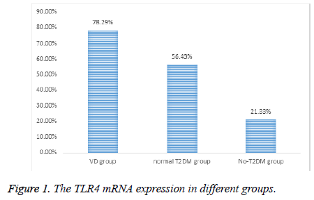
According to the reports of the technical department of Sino Biological Inc. the TLR4 mRNA expression was higher in VD group than other two groups (Figure 1).
CD4+/CD8+ ratio
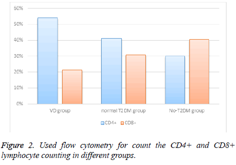
Using flow cytometry testing, the CD4+/CD8+ ratio of VD patients were increased significantly, almost tripling. Additional, the CD4+ counting was increasing, but CD8+ counting was decreasing (Figure 2).
Discussion
Globally as of 2010 it was estimated that there were 285 million people with T2DM making up about 90% of diabetes patients [12]. These risk factors of not only T2DM but also VD were analysed in our study, which were including overweight, age, residence, disease history (≥ 7 years), mental pressure, and contact without health management. So, health education must be emphasized in rural areas. And it need better health and food hygiene education to the blood glucose health management on early course of the T2DM disease. Since T2DM is typically a chronic disease, it associated with a tenyear- shorter life expectancy. This is partly due to a number of complications with which it is associated. This VD disease should be given enough attention from now-on. T2DM is a chronic metabolic disorders, one of the hazards is caused by chronic vascular complications, VD is one of the main causes of disability and death of neuroinflammation, which is inflammation of the nervous tissue [6,13].
On the other hand, cytokines and endothelium-related immune mediator expression levels increased significantly in the T2DM patients, according scientist’ reports [14,15]. As known, Toll- Like Receptor 4 (TLR4) is one of the most important recognizing antibody factors in the innate immune system. And TLR4 is an insulin resistance and its complications in the occurrence of important regulatory factors [16]. The molecular mechanism of cerebrovascular disease was confirmed in this study, about autoimmunity cytokines, TLR4. It found that TLR4 protein was at higher presence in VD group than None- VD group, which suggested that VD related with T2DM could increase the TLR4 presence related with tissue fibrosis. That was more likely to cause autoimmune neuritis. We might make a conclusion that VD is directly related with TLR4 increasing expression. And the result of CD4+/CD8+ ratio suggested that cell-mediated immunity is dominant during autoimmune process caused development of T2DM to VD. Through our study, it displayed TLR4 pathway on neuroinflammation conducted VD related T2DM. On the other field, more inflammatory factor should be tested in the future.
Acknowledgement
This study was supported by Project supported by Inner Mongolia Autonomous Region Natural Science Foundation (Grant No. 2015MS08136); Project supported by Inner Mongolia Autonomous Region Science and Technology Innovation and Guidance Foundation (Grant No. 201605).
References
- WHO. Diabetes Fact sheet N°312.
- National Institute of Diabetes and Digestive and Kidney Diseases. Diagnosis of diabetes and prediabetes 2016.
- Ghosh S, Trivedi S, Sanyal D, Modi KD, Kharb S. Teneligliptin real-world efficacy assessment of type 2 diabetes mellitus patients in India (Treat-India study). Diabetes Metab Syndr Obes 2016; 9: 347-353.
- Cunningham EL, McGuinness B, Herron B, Passmore AP. Dementia. Ulster Med J 2015; 84: 79-87.
- Qiang G, Wenzhai C, Huan Z, Yuxia Z, Dongdong Y, Sen Z, Qiu C. Effect of Sancaijiangtang on plasma nitric oxide and endothelin-1 levels in patients with type 2 diabetes mellitus and vascular dementia: a single-blind randomized controlled trial. J Tradit Chin Med 2015; 35: 375-380.
- Matei D, Popescu CD, Ignat B, Matei R. Autonomic dysfunction in type 2 diabetes mellitus with and without vascular dementia. J Neurol Sci 2013; 325: 6-9.
- Karantzoulis S, Galvin JE. Distinguishing Alzheimers disease from other major forms of dementia. Expert Rev Neurother 2011; 11: 1579-1591.
- Mahla RS, Reddy MC, Prasad DV, Kumar H. Sweeten PAMPs: Role of sugar complexed pamps in innate immunity and vaccine biology. Front Immunol 2013; 4: 248.
- Hansson GK, Edfeldt K. Toll to be paid at the gateway to the vessel wall. Arterioscler Thromb Vasc Biol 2005; 25: 1085-1087.
- Vijan S. In the clinic. Type 2 diabetes. Ann Intern Med 2010; 152: 31-35.
- Moroney JT, Bagiella E, Hachinski VC, Mölsä PK, Gustafson L, Brun A, Fischer P, Erkinjuntti T, Rosen W, Paik MC, Tatemichi TK, Desmond DW. Misclassification of dementia subtype using the Hachinski Ischemic Score: results of a meta-analysis of patients with pathologically verified dementias. Ann NY Acad Sci 1997; 826: 490-492.
- Singh A, Shenoy S, Sandhu JS. Prevalence of type 2 diabetes mellitus among urban Sikh population of Amritsar. Indian J Community Med 2016; 41: 263-267.
- Gendelman HE. Neural immunity: Friend or foe? J Neurovirol 2002; 8: 474-479.
- Matsuda T, Hisatsune T. Cholinergic modification of neurogenesis and gliosis improves the memory of AβPPswe/PSEN1dE9 Alzheimers disease model mice fed a high-fat diet. J Alzheimers Dis 2017; 56: 1-23.
- Yan T, Venkat P, Chopp M, Zacharek A, Ning R, Cui Y, Roberts C, Kuzmin-Nichols N, Sanberg CD, Chen J. Neurorestorative therapy of stroke in type 2 diabetes mellitus rats treated with human umbilical cord blood cells. Stroke 2015; 46: 2599-2606.
- Iori V, Iyer AM, Ravizza T, Beltrame L, Paracchini L, Marchini S, Cerovic M, Hill C, Ferrari M, Zucchetti M, Molteni M, Rossetti C, Brambilla R, Steve White H, Dincalci M, Aronica E, Vezzani A. Blockade of the IL-1R1/TLR4 pathway mediates disease-modification therapeutic effects in a model of acquired epilepsy. Neurobiol Dis 2016; 99: 12-23.

