ISSN: 0970-938X (Print) | 0976-1683 (Electronic)
Biomedical Research
An International Journal of Medical Sciences
Research Article - Biomedical Research (2017) Volume 28, Issue 6
Role of Rock 1 protein in non-small cell lung cancer
1Department of Clinical Pharmacy, Jilin University School of Pharmaceutical Sciences, Changchun, PR China
2Department of Cardiology and Chest Surgery, China-Japan Friendship Hospital of Jilin University, Changchun, PR China
3Yanbian University Medical College, Yanbian, PR China
4China Medical University, PR China
#These authors contributed equally to this work
- *Corresponding Author:
- Hua Xin
Department of Cardiology and Chest Surgery
China-Japan Friendship Hospital of Jilin University
Changchun, PR China
Accepted on November 04, 2016
The effects of Rock 1 in non-small cell lung cancer and the relationship between Rock 1 and non-small cell lung cancer were investigated in this study. Sixty cases of non-small cell lung cancer were investigated for the relationship between Rock 1 and non-small cell lung cancer. Expression of Rock 1 in non-small cell lung cancer was detected by histochemical staining and Real-time PCR. All the sixty cases had been diagnosed as non-small lung cancer by CT and HE staining. The expression of Rock 1 in nonsmall cell lung cancer was significantly higher than the adjacent tissues (p<0.05). Positive expression of Rock 1 was 73.33% in 60 cases. Among them, Positive expression of Rock 1 was 71.79% in squamous cell carcinoma of 39 cases. Positive expression of Rock 1 was 76.19% in adenocarcinoma of 21 cases. The expression of Rock1 may be positively related to the lung cancer, suggesting that Rock 1 play an important role in non-small cell lung cancer.
Keywords
Rock 1, NSCLC, Squamous cell carcinoma, Adenocarcinoma.
Introduction
It is well-known that lung cancer is currently the world incidence and mortality [1]. It is one of the worst malignant tumors, which seriously affects the health and survival of human beings [2]. In China, lung cancer has become the leading cause of death worldwide in 35 to 55 year of age [3]. According to World Health Organization data released in 2003, lung cancer was one of the most malignant tumors in the world [4,5]. Among them, non-small cell lung cancer is the most common in Lung cancer [6]. According to the report, the pathological types of non-small cell lung cancer include squamous cell carcinoma and adenocarcinoma [7]. In recent years, the investigation of the pathogenesis of non-small cell lung cancer has become a hot topic. Rock has a spiral structure, which is a main member of silk atmosphere acid/threonine [8]. In addition, it can be called Rho associated coiled coil forming protein kinase [9]. Recent studies had found Rock 1 was highly expressed in the tumor tissues such as non-small cell lung cancer, prostate cancer, liver cancer, ovarian cancer, and can promote the proliferation, invasion and metastasis of cancer cells [10-14]. Meanwhile, Rock 1 was closely related to the development of tumor, especially for non-small cell lung cancer [10]. The authors discussed the correlation between rock 1 protein and non-small cell lung cancer, and analyzed the expression of Rock 1 in non-small cell lung cancer. This study has laid a solid foundation for the targeted therapy of lung cancer.
Materials and Methods
Reagents and instruments
Rock 1 was purchased from Harling Biological Technology Co., Ltd (Shanghai, China). SP was purchased from Jinqiao Bio Technology Co., Ltd (Beijing, China). Biological Tissue Automatic Embedding Machine was purchased from taiva Technology Industrial Co., Ltd (Hubei, China). Olympus BX53 microscope was purchased from Leica Co., Ltd (German).
Clinical data
Paraffin-embedded tissue samples from 60 patients with clinical stage I-IIIA NSCLC were obtained from China-Japan Union Hospital of Jilin University from January 2013 to February 2014. There were 22 women and 38 men in 60 patients. The patient's age was from 36 to 73 years old. The average age was 58.78 years. All the samples were confirmed by histopathological examination. The results found that there were 39 cases of squamous cell carcinoma and 21 cases of adenocarcinoma. All of the samples were installed strictly in accordance with the requirements of medical ethics Association, and obtained the permission of patient's own or family member.
HE staining
Human lung cancer samples were fixed by 10% formaldehyde for 30 min and desiccated and embedded in paraffin. Xylene was used to remove paraffin for 10min, and 100%, 95%, 80% and 75% ethanol solution was used to replace xylene. Sections were washed with distilled water, stained with hematoxylin for 1 min. Dehydrated, transparent, mounting, camera.
Immunohistochemistry staining
Sections of human lung cancer tissue were sliced and roasted for 1 hour at 80°C. After deparaffinization and rehydration, antigen unmasking was performed by heating sections in 10 mm. The sections added to human Rock 1 antibody (1:200) at 4°C overnight, and washed three times with PBS and incubated with SP Kit (USA) for 20 min in light-shielded conditions. Sections were mounted with antifade reagents, covers lipped, and visualized microscopically.
Real-time PCR
Total RNA (mRNA) was extracted from Cancer and adjacent tissues according to the manufacturer's recommendations of TRIzol reagent (Invitrogen, Carlsbad, CA). 1 μg sample of total RNA was reverse transcribed to complementary DNA (cDNA) with oligo (dT) primers. We used amplified PCR fragments spanning different exons to prevent amplification of contaminated genomic DNA. The sense primer of ROCK1 was AAAATTGTGTGAGGAGGACATGG and the antisense primer was of TTCATCCCAACATTCTTGGATCT, which products were 279 bp in size. The house keeping gene GAPDH was used as an internal control. The sense primer was AGAAGGCTGGGGCTCATTTG and the antisense primer was AGGGGCCATCCACAGTC TTC, which products were 285 bp in size.
Positive judgment standard of rock 1 protein
Positive expression of Rock 1 is mainly brown or yellow particles. Expression of Rock 1 was analyzed by Plus Image- Pro image processing software.
Statistical analyses
All statistical analysis was performed using SPSS software version 15.0. The expression of Rock 1 was analyzed in triplicate. Numeric data were shown as means ± SD. Statistical significance between groups was analyzed by Student's t-test and one-way ANOVA. P<0.05 was considered statistically significant.
Results
CT detection
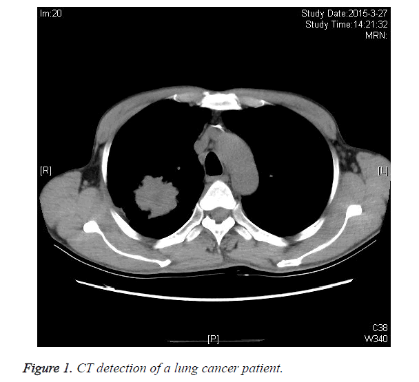
The patients were diagnosed by CT. Results showed that the patient was diagnosed with a small lung cancer. This diagnosis has laid a foundation for exploring the expression of Rock1 in lung cancer (Figure 1).
HE staining of squamous cell carcinoma
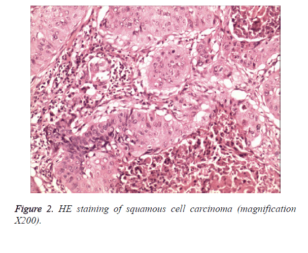
Sections of lung cancer were embedded in paraffin and stained, and then which sections were observed under the light microscope. The results showed that there was a part of the pearl or the partial horn within the nest of the cancer. The pathological mitotic figures were more obvious than the abnormal cells (Figure 2).
HE staining of adenocarcinoma
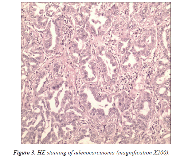
Sections of lung cancer were embedded in paraffin and stained, and then which sections were observed under the light microscope. The results showed that Adenocarcinoma cells arranged in adenoid or papillary. The atypia of cell was more obvious (Figure 3).
Expression of rock 1 protein in squamous cell carcinoma
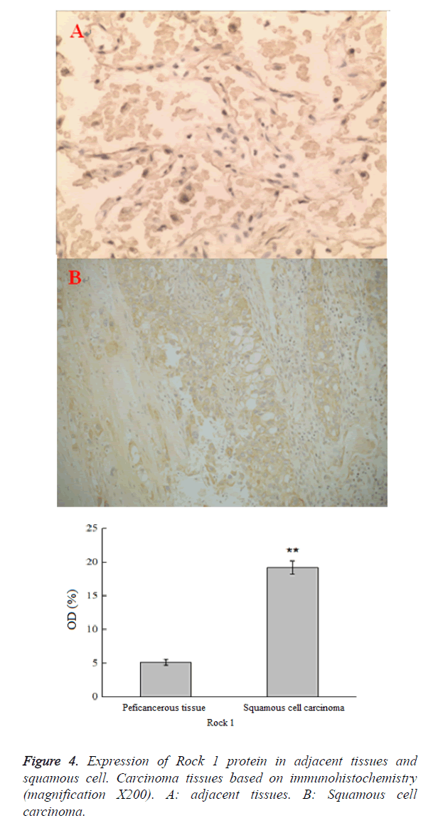
In this study, the expression of Rock 1 was determined in the squamous cell carcinoma tissues by immunohistochemistry. The results showed that there was small number of positive expression of Rock 1 in squamous cell carcinoma. In addition, the expression of Rock 1 in squamous cell carcinoma was significantly higher than that in the adjacent tissues (p<0.05). Meanwhile, the expression of Rock 1 in squamous cell carcinoma was brown and positive, which was mainly expressed in cytoplasm (Figure 4).
Expression of Rock 1 in lung adenocarcinoma
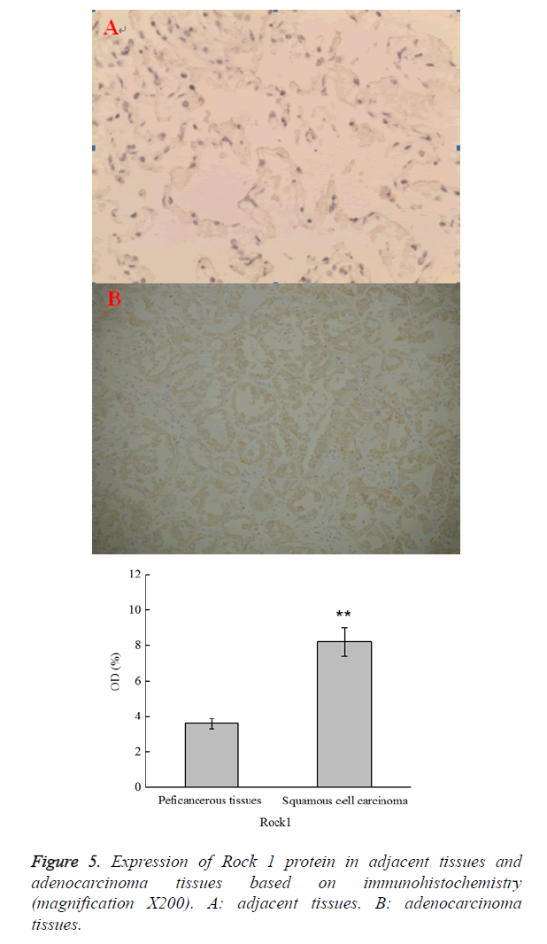
Expression of Rock 1 protein in the adenocarcinoma tissues was determined by immunohistochemistry. The results showed that there was small number of positive expression of Rock 1. The expression of Rock 1 in squamous cell carcinoma was significantly higher than that in the adjacent tissues (p<0.05).The expression of Rock 1 in adenocarcinoma was brown and positive, which was mainly expressed in cytoplasm (Figure 5).
Real-time PCR analysis
As shown in Table 1, the expression of Rock 1 mRNA in lung cancer was increased as compared with Paraneoplastic (P<0.01). These results showed Rock 1 have played an important role in squamous cell carcinoma and adenocarcinoma.
| Group | Squamous cell carcinoma | Adenocarcinoma |
|---|---|---|
| Paraneoplastic | 0.58 ± 0.01 | 0.42 ± 0.02 |
| Lung cancer | 1.23 ± 0.06** | 1.09 ± 0.05** |
| Lung cancer vs. Paraneoplastic P<0.01. | ||
Table 1. the radio of Rock I/GAPDH on Squamous cell carcinoma and Adenocarcinoma.
Correlation between Rock 1 expression and non small cell lung cancer
The positive expression rate of Rock 1 was 73.33% (44/60) in non-small cell lung cancer tissues. In addition, the positive expression rate of Rock 1 was 71.79% and 76.19% in squamous cell carcinoma (28/39) and adenocarcinoma (16/21), respectively (Table 2).
| N | Rock 1 expression | positive rate | ||
|---|---|---|---|---|
| Positive | Negative | |||
| Squamous cell carcinoma | 39 | 28 | 11 | 71.79% |
| Adenocarcinoma | 21 | 16 | 5 | 76.19% |
Table 2. Correlation between Rock 1 expression and non-small cell lung cancer.
Discussion
The development of tumor involves in many factors, such as activating of oncogene, invalidating of cancer suppressor genes and abnormal expressing of apoptosis genes [15]. Previous studies found that local invasion and metastasis of tumor cells included the complex biological processes of cell adhesion, the decomposition of the matrix around the cell, the invasion and metastasis [16]. In addition, the infiltration and metastasis of malignant tumor mainly depend on the migration of cells [17]. The change of cell morphology and adhesion is an important part of its ability to exercise and realize the infiltration and metastasis [18]. A large number of studies have shown that ROCK protein plays a significant role in the development of tumor [19]. Expression of ROCK was increased obviously in osteosarcoma, hepatocellular carcinoma, skin cancer tissue [20-21]. Effect of the substrate on many ROCK protein play a significant role in tumor development and related phenotypes, at the same time ROCK inhibitors can inhibit ability to migrate to a wide variety of tumor cell lines. ROCK protein involved in regulating tumor cell survival and apoptosis, invasion and metastasis of malignant tumor cells and other biological behavior [22].
Recent studies have found Rock 1 could regulate the relationship among the phosphate groups to proteins, intermediate metabolites, and remodeling the actin cytoskeleton, which affect the cell proliferation cycle and form of the phosphorylation reaction [11]. In this experiment, the results showed that the expression of Rock 1 in non-small cell lung cancer (squamous cell carcinoma and adenocarcinoma) tissues was significantly higher than that in the adjacent tissues, and the positive rate was 71.79% and 76.19%, respectively. Rock 1 is closely related to the development of tumor, especially in non-small cell lung cancer. With the development of lung cancer, the expression of Rock 1 protein increased obviously, indicating that expression of Rock 1 was closely related with non-small cell lung cancer. In a word, Rock 1 plays an important role in non-small cell lung cancer (adenocarcinoma and squamous cell carcinoma).
Conclusions
In conclusion, the current study demonstrated that the expression of Rock 1 was significantly increased in NSCLC tissue and the relationship between Rock 1 protein and non-small cell lung cancer.
References
- Wang Y, Liu Y, Yang B, Cao H, Yang CX, Ouyang W. Elevated expression of USP9X correlates with poor prognosis in human non-small cell lung cancer. J Thorac Dis 2015; 7: 672-679.
- Liu LZ, Fang J, Zhou Q, Hu X, Shi X, Jiang BH. Apigenin inhibits expression of vascular endothelial growth factor and angiogenesis in human lung cancer cells: implication of chemoprevention of lung cancer. Mol Pharmacol 2005; 68: 635-643.
- Chen W, Zheng R, Zhang S, Zhang S, Zhao P, Li G. Report of incidence and mortality in China cancer registries, 2009. Chin J Cancer Res 2013; 25: 10-21.
- Arrieta O, Quintana-Carrillo RH, Ahumada-Curiel G, Corona-Cruz JF, Correa-Acevedo E, Zinser-Sierra J. Medical care costs incurred by patients with smoking-related non-small cell lung cancer treated at the National Cancer Institute of Mexico. Tob Induc Dis 2015; 12: 25.
- Henzler T, Goldstraw P, Wenz F, Pirker R, Weder W, Apfaltrer P. Perspectives of novel imaging techniques for staging, therapy response assessment, and monitoring of surveillance in lung cancer: summary of the Dresden 2013 Post WCLC-IASLC State-of-the-Art Imaging Workshop. J Thorac Oncol 2015; 10: 237-249.
- Yu Z, Lu H, Si H, Liu S, Li X, Gao C. A Highly Efficient Gene Expression Programming (GEP) Model for Auxiliary Diagnosis of Small Cell Lung Cancer. PLoS One 2015; 10: e0125517.
- Nizzoli R, Tiseo M, Gelsomino F, Bartolotti M, Majori M, Ferrari L. Accuracy of fine needle aspiration cytology in the pathological typing of non-small cell lung cancer. J Thorac Oncol 2011; 6: 489-493.
- Chen HC, Chang JP, Chang TH, Lin YS, Huang YK, Pan KL. Enhanced expression of ROCK in left atrial myocytes of mitral regurgitation: a potential mechanism of myolysis. BMC Cardiovasc Disord 2015; 15: 33.
- Sawada N, Liao JK. Rho/Rho-associated coiled-coil forming kinase pathway as therapeutic targets for statins in atherosclerosis. Antioxid Redox Signal 2014; 20: 1251-1267.
- Kang CG, Han HJ, Lee HJ, Kim SH, Lee EO. Rho-associated kinase signaling is required for osteopontin-induced cell invasion through inactivating cofilin in human non-small cell lung cancer cell lines. Bioorg Med Chem Lett 2015, 25: 1956-1960.
- Zhang C, Zhang S, Zhang Z, He J, Xu Y, Liu S. ROCK has a crucial role in regulating prostate tumor growth through interaction with c-Myc. Oncogene 2014; 33: 5582-5591.
- Au SL, Wong CC, Lee JM, Wong CM, Ng IO. EZH2-mediated H3K27me3 is involved in Epigenetic repression of deleted in liver cancer 1 in human cancers. PLoS One 2013; 8: e68226.
- Ohta T, Takahashi T, Shibuya T, Amita M, Henmi N, Takahashi K. Inhibition of the Rho/ROCK pathway enhances the efficacy of cisplatin through the blockage of hypoxia-inducible factor-1α in human ovarian cancer cells. Cancer Biol Ther 2012; 13: 25-33.
- Han ZQ, Hong ZY, Hu CX, Hu Y, Chen CH, Lu YP. Effect of blocking of ROCK-1, an effector of small G protein Rho, on the malignant behavior of ovarian tumor cells in vitro. Zhonghua Zhong Liu Za Zhi 2007; 29: 723-727.
- Zhang B, Chen B, Wu T, Xuan Z, Zhu X, Chen R: Estimating developmental states of tumors and normal tissues using a linear time-ordered model. BMC Bioinformatics 2011; 12: 53.
- Gupta SC, Kim JH, Prasad S, Aggarwal BB. Regulation of survival, proliferation, invasion, angiogenesis, and metastasis of tumor cells through modulation of inflammatory pathways by nutraceuticals. Cancer Metastasis Rev 2010; 29: 405-434.
- Raore B, Schniederjan M, Prabhu R, Brat DJ, Shu HK, Olson JJ. Metastasis infiltration: an investigation of the postoperative brain-tumor interface. Int J Radiat Oncol Biol Phys 2011; 81: 1075-1080.
- Li S, Braverman R, Li H, Vass WC, Lowy DR, DeClue JE. Regulation of cell morphology and adhesion by the tuberous sclerosis complex (TSC1/2) gene products in human kidney epithelial cells through increased E-cadherin/beta- catenin activity. Mol Carcinog 2003; 37: 98-109.
- Yousif MH, Makki B, El-Hashim AZ, Akhtar S, Benter IF. Chronic treatment with Ang-(1-7) reverses abnormal reactivity in the corpus cavernosum and normalizes diabetes-induced changes in the protein levels of ACE, ACE2, ROCK1, ROCK2 and omega-hydroxylase in a rat model of type 1 diabetes. J Diabetes 2014; 2014:142154.
- Liu X, Choy E, Hornicek FJ, Yang S, Yang C, Harmon D. ROCK1 as a potential therapeutic target in osteosarcoma. J Orthop Res 2011; 29: 1259-1266.
- Wong CC, Wong CM, Tung EK, Man K, Ng IO. Rho-kinase 2 is frequently overexpressed in hepatocellular carcinoma and involved in tumor invasion. Hepatology 2009; 49: 1583-1594.
- Samuel MS, Lopez JI, McGhee EJ, Croft DR, Strachan D, Timpson P. Actomyosin-mediated cellular tension drives increased tissue stiffness and β-catenin activation to induce epidermal hyperplasia and tumor growth. Cancer Cell 2011; 19: 776- 791.
