ISSN: 0970-938X (Print) | 0976-1683 (Electronic)
Biomedical Research
An International Journal of Medical Sciences
Research Article - Biomedical Research (2017) Artificial Intelligent Techniques for Bio Medical Signal Processing: Edition-I
Research on the application of the fusion of medical imaging technology in the treatment of the knee injury in competitive sports
1Basketball Teaching and Research Office of xi'an Physical Education University, Shaanxi, PR China
2School of Computer Science & Technology, Xi`an University of Posts and Telecommunications, Shanxi, PR China
- *Corresponding Author:
- Youping He
Basketball Teaching and Research Office
Xi'an Physical Education University, PR China
Accepted on January 09, 2017
Sports athletes easily get joint damage due to the high intensity of physical training and exertion. The treatment of joint injury caused by physical exercise is very important for competitive sports. Joint composition structure is complex and the knee joint is considered as an example in the full text and the application of CT imaging technology in the diagnosis and treatment of knee joint was studied, which a certain clinical significance for the treatment of joint injury of athletes. In this work it is proposed to establish a unified registration system and statistical coordinate system to achieve image fusion. Secondly, the optimization design of the fusion image algorithm based on the virtual projection CT data was carried out and the image was converted into the same coordinate system. Carry out the final image fusion. The X-ray image and CT image data were combined to obtain the absolute coordinates of the registration position and then the image was fused. Finally, the fusion medical imaging technology was applied in the diagnosis and treatment of knee joint injury. It can be seen through data collection and analysis on 21 cases of patients that the integration of medical imaging technology can diagnosis knee injury accurately, thereby reducing unnecessary surgery and improving the rehabilitation effect of patients.
Keywords
Fused image, Knee joint, Injury, CT, X-ray image.
Introduction
The injury of the joint is higher because of the high intensity training and competition in athletic sports. A lot of excellent athletic athletes influence the development of their sports career. For example, the basketball player Liu Yudong and Yao Ming finally retired in recent years, which is more or less with the knee injury. Gymnast Chen Yibing retired with helpless because of knee meniscus injury and tennis player Li Na retired with related knee [1]. Whether it is the track and field events or other sports, the knee joint and athletes are closely related to the sport in competitive sports. The knee joint injury and medical image diagnosis was considered as the research object, and then the results of the study were applied to the joint injury treatment of sports athletes [2]. Use medical imaging technology for the diagnosis of the knee injury to help the subsequent treatment and reduce the pain of patients [3]. However, a single medical imaging accuracy rate was not high on the condition of the patient assessment, which not only affected the timely treatment of patients, but also may be to the effective treatment due to the wrong choice treatment because the knee joint structure is more complex [4]. The common medical imaging technology of knee joint is CT and X-ray image technology. Integrating the two can make full use of each other's advantages and improve the accuracy of medical image evaluation, which has a positive clinical significance for the treatment of knee injury.
Related Theoretical Research
Injury of knee joint: As a common injury in sports, the influence of the knee injury on the athletes is great, and the competitive sports athletes will have the situation of the knee injury inevitably (Figure 1). The injury of meniscus and ligament is more in these injuries. The composition of the knee joint consists of two parts of tibia and patella, which can complete flexion, rotation, and sliding action, and main structures included bone, meniscus, cartilage, ligament, synovium and synovial fluid, fat pad [5]. Whether athletes or general sports, they should pay attention to the protection of the knee joint and the treatment of knee injury. The diagnosis of knee joint used CT and X-ray imaging commonly, which was able to obtain a higher resolution and diagnostic accuracy. Combined with medical imaging technology was also helpful for clinical treatment in the treatment of knee injury [6]. Knee joint injury included ligament injury, meniscus injury and complications mainly. As the most prone to injury in the knee joint, the meniscus injury was the condition of the loss of the knee joint in most sports athletes. The meniscus is between the inside and outside of the femur and the tibial plateau, and the medial meniscus and the lateral meniscus were presented "C" and "O" [7]. Normal meniscus was composed of fibrous cartilage, and the injured meniscus was invaded by the joint fluid, so the proton density has been changed and the proton cyclotron rate decreases. Ligaments have a stable effect on the knee joint, including anterior and anterior cruciate ligament, collateral ligament [8]. The hydrogen proton is fixed on a dense grid of multiple units because of the normal ligament. A low signal that is difficult to be imaged on the sequence when use of MRI imaging technology [9]. When the ligament is injured, because the ligament appears edema, hematoma, and so on, there will appear the situation of local high signal. MRI imaging technique was observed in the ligament of the low signal walking area is not continuous [10]. In addition, there are a variety of complications, such as discoid meniscus, plica syndrome, exfoliative cartilage and synovitis. But more common is the ligament injury and meniscus injury in sports [11].

Figure 1: Knee joint injury in sports.
Medical image fusion technology: Medical imaging technology has developed rapidly in recent years, which provides sufficient reference image for the diagnosis and treatment of various diseases, and helps doctors with clinical symptoms and other tests to determine the condition of patients [12]. However, there are some problems in the image obtained by the single medical image method, such as the data obtained is not complete, the results are not accurate enough, the image fuzzy, etc. In addition to the continuous development of existing medical imaging technology, people still in full research of medical image fusion technology in order to solve these problems [13]. Medical image fusion technology is two or more than two kinds of medical imaging technology for the scientific comprehensive, which gives full play to the advantages of each other with two of the acquired data, cross reference verification, and then reconstruct the image more clear, accurate and reliable. The knee joint was easy to be injured in motion and the traditional medical imaging technology was difficult to study the motion state of the knee joint accurately. Therefore, it was necessary to make more accurate diagnosis of the injury of the knee joint in the treatment process [14]. The combination of X-ray image technology and CT imaging technology can fully combine the advantages of the two. The image reconstruction of the knee joint was more accurate.
Computed radiography was one of the commonly used imaging techniques at present with high spatial resolution, dynamic range of the advantages, especially for the diameter of not less than 2 mm. The contrast is not more than 1% objects observed and the image effect was better [15]. The radiation dose produced by the image was low and the influence on the human body was small when using the computer X-ray photography technology. Use computer radiography technology can improve the image details, realize image amplification and digital subtraction function, decrease noise reduction while adjusting the contrast of the image and show the characteristic information of many other imaging techniques difficult to observe. However, this technology also had some shortcomings. Because all the structure was a twodimensional projection superposition in the human body, so the three-dimensional structure was not clear with the lack of three-dimensional information [16]. Its working principle diagram was shown in Figure 2.

Figure 2: Principle of computer X-ray photography.
CT image technology was the use of human body structure on the absorption of different X-ray images to obtain the structure of the human body, which can fully reflect the distribution of different tissues of proton density, thus forming a full range of images from different angles [17]. The absorption coefficient of the bone and the larger organization will show a clear image due to the different tissues of the body to the X-ray absorption coefficient is very different. The corresponding spatial resolution is higher, but the soft tissue has less absorption coefficient, so the imaging effect is poor. And the content of the proton in the tissue is proportional to the signal intensity of the imaging. If the difference between the X-ray absorption coefficient of the tissue was larger, the imaging effect was more clear [18]. The working principle of CT image technology is shown in Figure 3. In summary, whether CT technology or computer radiography technology, there are still some problems. The medical image registration and fusion technology based on a variety of medical imaging technology has become the development direction of more and more medical imaging problems in recent years. Fusion of medical imaging technology was first started in 1970s, and now has achieved considerable success after decades of rapid development. 2D/3D image registration can be used to combine the two together. Play each other's data advantages and develop more suitable for clinical practice algorithm and application.

Figure 3: Working principle of CT image technology.
Application of Fusion Medical Imaging Technology in the Diagnosis and Treatment of Knee Joint Injury
Fusion medical imaging technology: We must first establish a unified registration system to realize the fusion of different image technology images. The establishment of registration system was completed by establishing the 2D/3D registration principle in this paper [19]. The process of optimization of multi parameters can be realized by iteration because there are many parameters in the process of spatial transformation. The registration method was based on gray feature or geometric feature. The former was based on DRR and X-ray image, and the latter was based on the projection profile [20]. The similarity measure function was constructed according to the DRR image and the image of the X-ray image and the spatial position parameters of the two images. It was able to carry out the fusion of two kinds of images and complete the registration between the two through the calculation of the optimal solution. Bhatnagar et al. and Xu extensively proposed fusion techniques and can be found in current literature [21,22]. Further, calculate the spatial position of the two kinds of image changes and conduct the integration of the various parts of the space position. The 3D motion data and the steps obtained are as in Figure 4.

Figure 4: 2D/3D medical image registration principle.
As the first step of medical image fusion, we must first set the three relations of point light source, body data and the position of the receiving screen to obtain image registration. The image can be improved by changing the position in the process of 2D/3D image registration. The same image can be obtained only if the thickness of the object was not regular and only in the case of the source that the receiver and the position of the object. So it is necessary to set up the point light source and the virtual receiver (Figure 5). Set the calibration method used in the calibration board is a hollow square copper wire and the copper wire width is 0.254 mm, which is more convenient to calculate and can fit the more accurate straight line. Camera calibration algorithm has been quite mature. The obtained images are processed in the calibration toolbox camera based on the use of Matlab language. Its specific process is as in Figure 6. CT device itself has been identified in the coordinate system. Its coordinate origin was generally located in the lower left corner and X, Y, Z axis were in accordance with the rules of the right hand screw. Set it to CT original coordinate system. It was time to complete the image fusion and it was necessary to reduce all the coordinate system. That is the registration coordinates. Determine the coordinate system to reduce the amount of data involved in the fusion process as much as possible and optimize the relevant calculation process. CT data was more likely to be reconstructed after translation or rotation. The coordinate system and parameters were set as shown in Figure 7.

Figure 5: Process flow chart for the fusion of medical imaging technology.

Figure 6: Image shooting condition calibration process.

Figure 7: Coordinate system and corresponding parameter settings.
The second is to realize the unification of pixels. We must consider the real scene, which can obtain the fused image based on the X-ray image when setting the parameters. Image fusion images are from the CT equipment and DR equipment and the basic unit of the image is pixels. However, it needs to measure the unity due to their size and resolution of the pixel is not consistent. CT get data images are founder and Pixels are generally 512 × 512. The specific size is determined by the image reconstruction. The final voxel size is x layer thickness combined with the layer thickness of the scan. The image information captured by the DR device is determined by the original setting and is stored in a DICOM format. A new pixel image is calculated according to the relationship between the source and the screen in the calibration system:
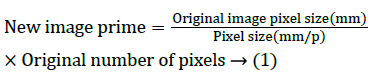
Three kinds of pixel data are completed, which are the transformation of the source screen position relation, the transform of CT data and the change of the X-ray image through the 2D/3D registration process. The process of its transformation is shown in Figure 8. The transformation of the source and the position of the screen are to calculate the source screen position, which is obtained by the intrinsic relationship between the pixel and the pixel size so as to obtain the relationship between the number of pixels in the virtual environment. The CT image pixel and registration system of the pixel is set to become the relationship between the same proportion of 1:1. Conduct data set in advance before each input CT. The original pixel physical size of X-ray image converts to the registration of the main program to match the number of pixels in the transformation.

Figure 8: Unified unit of parameter conversion.
Optimization design of image fusion algorithm: The gray value of CT scan in the image is also different because of the different density of bone, skin, muscle, blood and so on in human body tissue. The overall CT data can be seen as a virtual reconstruction of objects. CT imaging technology and X-ray image technology were conducted fusion and reconstruction, which was used ray tracing algorithm. Virtual light source is emitted from the virtual point light source through the CT data acquired by the scanning, and then received and imaged on the virtual image. Similar to the principle of CT scanning, the formation of virtual image is also a continuous attenuation of virtual light. And it is mapped to the 1-255 gray scale after the calculation is completed, which can be used to display the pseudo visible light density and finally the image is reconstructed. It is to be absorbed and scattered in the path of X-ray emission because of the absorption and scattering of the beam. The attenuation coefficient of the X ray is μ, the distance of the ray passes through is d, the ray intensity is I0, and the attenuation of the X-ray can be calculated:
 → (2)
→ (2)
Correspondingly, the distance and the linear attenuation coefficient di are μi and respectively, which can be got through by the type (2):
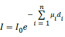 → (3)
→ (3)
It can be deduced through the nearest neighbor method to calculate the virtual X-ray attenuation:
 → (4)
→ (4)
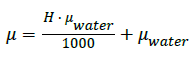
The conversion factor is used to convert it into:
 (5)
(5)
Energy attenuation is largely ignored in the air in formula (5), and thus. F=0
The volume data obtained in the three coordinate directions are not consistent and the pixels are generally square when the ray tracing algorithm is used, so the small rectangular body with different volume can be formed. As a result, the layer thickness of the CT data is reduced and the image can be acquired more clearly when data is reconstructed. When the CT data is interpolated by the adjacent interpolation method, the effect is better.
Coordinate transformation is the transformation of the rigid
body when the image is fused with time. The distance between
the one or two points of the rigid body is not changed with the
change of the space position and the distance between two
points is changed. Therefore, the transformation of the rigid
body is divided into rotation, translation, and specular
reflection. The position and direction change with the
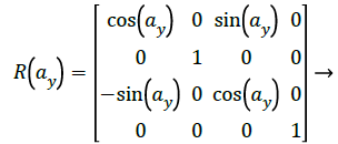
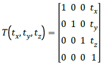
parameter vector 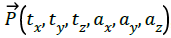 , which was expressed
along the X axis, Y axis and the Z axis of the continuous
rotation respectively. These six degrees of freedom parameter matrix form is:
, which was expressed
along the X axis, Y axis and the Z axis of the continuous
rotation respectively. These six degrees of freedom parameter matrix form is: 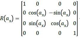 → (6)
→ (6)
 (7)
(7)
 → (8)
→ (8)
 → (9)
→ (9)
 → (10)
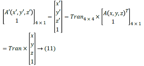
Set a coordinate point as A and the three-dimensional coordinates of the point is A’ after the above conversion in the registration coordinate system of the reconstructed image. The relationship between the two is:
The X-ray image is used as the reference image, and the final fused DRR image is used as the floating image. The reconstructed image is obtained after changing the angle and position of the virtual perspective and position of the CT body data. Then it compares the similarity with the original X image until the maximum value is obtained. The difference between the two is the smallest and the similarity measure can be carried out with the same mode imaging for the two images in the same imaging mode. Assume the reference image is R(i,j), the floating image is F(i,j), the image is Ft ’(i,j), and finally the objective function is optimized:
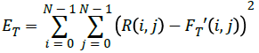 → (12)
Multiple extreme points may occur in the process of solving the minimum value of ET. Therefore, human intervention is needed to get the best results.
Convert to the centroid position and converted again to the origin of software in the coordinate system in the use of Raindrop Geomagic to CT data conversion origin. The 3D point cloud file can be converted into the software coordinate system when the coordinate system which is consistent with the 2D/3D registration procedure is adopted. The unified process of coordinate system is shown in Figure 9.

Figure 9: Unity of coordinate system.
Application of fusion medical imaging technology in the diagnosis and treatment of knee joint injury: The athletes will take some protective measures in the process of movement in order to reduce knee injury, such as early warm-up exercise and knee pads. However, it is inevitable that there will be an accident in the movement resulting in the injury of the knee joint. The knee injury may involve the bones, ligaments, joints, cartilage and soft tissue and so on, so once the knee injury, the damage may occur more. Conventional medical imaging technology is difficult to accurately diagnose the injury of the knee joint, which may affect the effect of the treatment directly. We can reconstruct the overall spatial structure of the knee joint by using the fusion technology of medical image registration, and obtain more accurate information of the knee joint by using the technique, which is helpful for the treatment of joint injury.
The three-dimensional structure of the knee joint can be obtained by the fusion technology of CT and X-ray image. Each bone of the knee joint can be considered as a rigid body. It is needed to carry out a continuous space positioning to complete the image fusion. CT scanning equipment used is the second generation dual source spiral CT. Image size and pixel should be set according to the specific conditions of the shot. The knee joint was placed in the CT equipment for thin layer scanning. First of all, conduct the X-ray image when the DR shooting. The device source screen is arranged at a distance of about 100 cm, and the bottom pad is padded with some foam boards to keep the position of the DR equipment to be the same. Always keep the knee in the process of shooting. Complete the laser 3D scanning and X-ray image after each shift position or position. You should be clear the relationship between the two in the preservation of and take a total of 8 images of the film before and after.
The X-ray image and CT image data were integrated to obtain the absolute coordinates of the registration position by using the optimization calculation method. Select the first DR image and compare the back of the image with the contrast so as to calculate the CT data between the images of the space transformation. Conduct image fusion and reconstruction and the reconstructed images and real X images are interpolated and processed by image processing to analyze the effect of image fusion ultimately from a visual angle according to the registration result after obtaining the CT sequence image. Mosaic image is the two images of the same size were processed into 256 uniform square areas. Splicing a part of the two images and the specific splicing process is shown in Figure 10.

Figure 10: Schematic diagram of mosaic image.
Application Analysis
Materials and methods: X-ray image technology and CT image technology of the fusion image technology were used. Collect the related data of patients with knee joint injury during the period of February -2015 in a hospital in October 2014. Among them, 13 were male and 8 were female. The ratio of male to female was 1.625:1. The patient's age range was 16-41 years old. The mean age was 30.4 years old, and the course of disease was 1 days -6 months. The main clinical symptoms and signs are: joint pain, local swelling, obvious injury, the degree of activity is restricted, etc. All patients were admitted to hospital for routine MRI examination and imaging examination using the fusion of medical imaging technology. Choose whether to need to carry on the operation and what kind of operation according to the patient's condition.
The equipment used in the main CT equipment and X-ray images of the DR devices. The specific parameters are as follows: CT equipment is Siemens 2008G with the two generation dual source spiral CT, voltage 80KV and scanning time is 1000 ms, 512 * 512 pixel image. The equipment used in the X-ray image is AG Siemens, DE-80333, voltage 60 KV, the tube voltage is 450 mA. The image size is according to the situation, the exposure time is 10 ms.
All patients underwent CT scan and X-ray scan. Then the calibration procedure is used to process the image, and the absolute coordinates of the registration position are obtained. Then the software is used to carry out the registration of point cloud registration, and the registration parameters are obtained by comparing with the reference. Finally, conduct image mosaic. Conduct comprehensive analysis of the injury of the knee by using the image of the fused image to guide the clinical diagnosis and treatment of the knee joint.
Result analysis: The results of the examination by the fusion of medical imaging technology are as follows:
(1) There are 3 cases level 1 half-moon damage, among them lateral meniscus is in 1 cases and medial meniscus injury is in 2 cases. There are 4 cases 2 level meniscus injury, among them lateral meniscus is in 2 cases and medial meniscus injury is in 2 cases. There are 4 cases 3 level meniscus injury, among them lateral meniscus is in 3 cases and medial meniscus injury is in 1 cases. There were 9 cases with the imaging diagnosis and the accuracy rate was 81.82% in the subsequent surgical treatment.
(2) There were 9 cases of ligament injury in 21 cases, including 3 cases of anterior cruciate ligament injury and 6 cases of posterior cruciate ligament injury. The performance of ligament injury and imaging technology has been the performance of 8 cases and the diagnostic accuracy rate of 88.89% in the treatment of ligament injury.
(3) There are 2 cases of combined disk meniscus, 1 cases of peeling off and 3 cases of joint effusion accompanied by the knee joint injury of the combined symptoms.
In summary, using the fusion of medical imaging technology can diagnose the knee injury accurately, which has a very important clinical significance for the diagnosis and treatment of the whole knee joint injury. We can improve the accuracy rate of initial diagnosis, reduce unnecessary surgical treatment, reduce the pain of patients and improve the patient's rehabilitation effect by using this technology.
Conclusion
Joint injury in sports activities is one of the serious threats to the health of the athletes for competitive athletes. Taking medical imaging technology is the main method of clinical diagnosis of joint injury. Compared with the traditional single medical imaging technology, medical imaging technology can be used to get more clear images to make a more accurate assessment of the joint injury in order to improve the accuracy of diagnosis. The combination of X-ray image technology and CT imaging technology can give full play to the advantages of each other. Taking the diagnosis of the knee joint injury as an example, conduct the realization of the coordinates of the two images through the establishment of a unified registration system. Conduct image reconstruction of CT data by using virtual projection. Conduct image stitching and generate the fusion image ultimately through the optimization of the fusion image algorithm design. The accurate rate of diagnosis was 81.82% month injury and the diagnosis of ligament injury and the accuracy rate was 88.89% through the collection and analysis of hospital patients with knee joint injury data and using imaging fusion of medical imaging technology. Fusion of medical imaging technology can diagnose the knee joint injury accurately, which has a certain clinical significance.
References
- Wu CL, Jain D, McNeill JN, Little D, Anderson JA. Dietary fatty acid content regulates wound repair and the pathogenesis of osteoarthritis following joint injury. Ann Rheum Dis 2015; 74: 2076-2083.
- Whittaker JL. Evidence of ealy post-traumatic ostearthritis and other negative health outcomes 3-10 years following knee joint injury in youth sport. World Confederation for Physical Therapy 2015.
- Kras JV, Weisshaar CL, Pall PS, Winkelstein BA. Pain from intra-articular NGF or joint injury in the rat requires contributions from peptidergic joint afferents. Neurosci Lett 2015; 604: 193-198.
- Balke M, Schneider MM, Shafizadeh S. Current state of treatment of acute acromioclavicular joint injuries in Germany: is there a difference between specialists and non-specialists? A survey of German trauma and orthopaedic departments. Knee Surgery Sports Traumatol Arthroscopy 2015; 23: 1447-1452.
- Laprade RF, Smith SD, Wilson KJ. Quantification of functional brace forces for posterior cruciate ligament injuries on the knee joint: an in vivo investigation. Knee Surgery Sports Traumatol Arthroscopy 2015; 23:3070-3076.
- Whittaker J, Woodhouse L, Nettel-Aguirre A. Evidence of early post-traumatic osteoarthritis and other negative health outcomes 3-10 years following knee joint injury in youth sport. Osteoarthritis Cartilage 2015; 23:e1662-e1663.
- Sahin N, Bianco A, Patti A. Evaluation of knee joint proprioception and balance of young female volleyball players: a pilot study. J Physical Ther Sci 2015; 27:437-440.
- Nomura M, Iwasawa H, Sakitani N. Spinal cord injury leads to region specific tidemark advancement in articular cartilage of the knee joint in rats. Physiotherapy 2015; 101:e1103-e1104.
- Yoo JH, Lee JH, Chang CB. Pure Varus Injury to the Knee Joint. Clin Orthop Surg 2015; 7: 269-274.
- Abaza H. Effect of balance training, plus black seeds capsules on knee joint injuries, balance and performance. J Am Sci 2015; 11: 136-139.
- Junge T, Larsen LR, Juul-Kristensen B, Wedderkopp N. The extent and risk of knee injuries in children aged 9–14 with Generalised Joint Hypermobility and knee joint hypermobility - the CHAMPS-study Denmark. Bmc Musculoskeletal Disord 2015; 16:1-11.
- Liu S, Zhang T, Li H. Medical image fusion based on nuclear norm minimization. Int J Imaging Syst Technol 2015; 25:310-316.
- Clingman B, Miller T, Oraevsky A. Opto-acoustic image fusion technology for diagnostic breast imaging in a feasibility study. SPIE Med Imaging2015.
- Pu T, Ni G. Contrast-Based Image Fusion Using the Discrete Wavelet Transform.Opt Eng 2015; 39: 2075-2082.
- Bhavana V. Multi-Modality Medical Image Fusion- A Survey. Int J EngRes Technol 2015; 2015:778-781.
- Mourão A, Martins F, Magalhães J. Multimodal medical information retrieval with unsupervised rank fusion. Comput Med Imaging Graphics 2015; 39:35-45.
- Foncubiertarodríguez A, Müller H. Medical case-based retrieval: integrating query MeSH terms for query-adaptive multi-modal fusion. SPIE Med Imaging2015.
- Lo RC, Huang WL, Hsiao YL. Performance evaluation of the section of 3D reconstruction based on different PET/CT image fusion sequence. Technol Health Care 2015.
- Alshawi TA, El-Samie FEA, Alshebeili SA. Magnetic resonance and computed tomography image fusion using bidimensional empirical mode decomposition. IEEE Global Conference on Signal and Information Processing 2015.
- Liu S, Zhang T, Li H. Medical image fusion based on nuclear norm minimization. Int J Imaging SystTechnol 2015; 25:310-316.
- Bhatnagar G, Wu QJ, Liu Z. Directive contrast based multimodal medical image fusion in NSCT domain. IEEE Transact Multimedia 2013; 15: 1014-1024.
- Xu Z. Medical image fusion using multi-level local extrema. Information Fusion 2014; 19: 38-48.