ISSN: 0970-938X (Print) | 0976-1683 (Electronic)
Biomedical Research
An International Journal of Medical Sciences
Research Article - Biomedical Research (2017) Volume 28, Issue 11
MicroRNA-320 inhibits insulin resistance in patients with PCOS through regulating ERK1/2 signaling pathway
1Department of Obstetrics and Gynecology, the Second Affiliated Hospital of Zhengzhou University, Zhengzhou, Henan, PR China
2Department of Reproductive Medicine, the Second Affiliated Hospital of Zhengzhou University, Zhengzhou, Henan, PR China
- *Corresponding Author:
- Wen-Na Yuan
Department of Obstetrics and Gynecology
The Second Affiliated Hospital of Zhengzhou University, PR China
Accepted date: March 28, 2017
Objective: This study aimed to investigate the role of microRNA-320 in Insulin Resistance (IR) of patients with Polycystic Ovary Syndrome (PCOS) and its regulatory mechanism.
Methods: Ovarian tissues in 20 patients with PCOS and IR (PCOS-IR group) and 20 normal persons (control group) were collected. The total RNA was extracted from each ovarian tissue. The differential microRNA-320 expression in patients of PCOS-IR group and control group were verified by RT-PCR. TargetScan and miRanda were used to predict the possible specific binding target sites with microRNA-320. The dual-luciferase reporter gene method was used to predict the specific binding target sites of 3’ UTR. The regulatory pathways of ovarian tissues in patients of PCOS-IR group and control group were predicted by the Western blot method.
Results: The microRNA-320 expression in the ovarian tissue in the PCOS–IR group was significantly lower than that of the control group. The IRS-1 specific binding target sites with microRNA-320 were screened by the bioinformatic software and dual-luciferase reporter gene method. Moreover, compared with the control group, the expression of ERK1/2 was significantly up-regulated in PCOS group.
Conclusion: MicroRNA-320 could inhibit IR in patients with PCOS through IRS-1 regulating ERK1/2 signaling pathway.
Keywords
Microrna-320, PCOS, Insulin resistance, ERK1/2.
Introduction
Polycystic Ovary Syndrome (PCOS) refers to a series of syndromes, including bilateral ovarian polycystic changes, chronic anovulation, or rare ovulation, accompanied by infertility, hirsutism, acne, and menstrual disorders [1]. The disease can seriously affect fertility of patients. The clinical features of PCOS include hyperandrogenism and absence or sparse ovulation. PCOS is the most common endocrine disease in 5%-10% of women of childbearing age [2]. At present, PCOS is considered to be an endocrine and metabolic disease characterized by Insulin Resistance (IR). The hyperinsulinemia secondary to IR plays an important role in androgen excess. MicroRNA is a kind of small-molecule single-stranded noncoding RNA that binds with mRNA specific target sequence and inhibits the expression of target gene, to regulate the posttranslational expression regulation and the development and physiological function of tissue and organ [3,4]. The present studies have showed that microRNA-21, microRNA-27b, microRNA-103, and microRNA-155 are involved in the regulation of follicular development, androgen synthesis, and insulin sensitivity-related signaling pathways, which may provide a new target for the diagnosis and treatment of PCOS [5]. The purpose of the present study is to investigate the differential expression of microRNA-320 in patients with PCOS and patients without PCOS and its role in the pathophysiological mechanism.
Materials and Methods
Reagents and instruments
The QuikChange® site-specific mutagenesis kit was purchased from Stratagene Corporation, Shanghai. The lipofection transfection reagent Lipofectamine® 3000 was purchased from Invitrogen Corporation. The Dulbecco’s modified Eagle’s medium, fetal bovine serum and trypsin were purchased from Gibco. The CCK-8 kit was purchased from Biyuntian Packing Product, Co., Ltd., Shanghai. The clean bench and carbon dioxide incubator were purchased from Thermo Fisher Scientific, USA. The P-ERK1/2 and ERK1/2 were purchased from CST, USA. The Horseradish Peroxidase (HRP)-labeled secondary antibody was purchased from Boshide Company, Wuhan.
Tissue specimen
The ovarian tissues in 20 cases of patients with PCOS and IR (PCOS-IR group) and 20 cases of normal persons (control group) were collected. The ovarian tissue was flushed with phosphate-buffered saline, cut into 0.1 cm3 with ophthalmic scissors, and preserved in liquid nitrogen.
Cell sources
The human embryonic kidney 293T cell was purchased from the Shanghai Institutes for Biological Sciences of the Chinese Academy of Sciences.
RNA extraction, inversion, and RT-PCR detection
RNA from the ovarian tissue was extracted and quantified by the TRizol method. The extracted RNA was reversely transcribed into cDNA using the AMV reverse transcriptase. The cDNA was used as the template, and RT-PCR was performed. The microRNA-93 was detected by RT-PCR. The primer was synthesized by Shanghai Sangon Biotechnology Company Ltd., and the synthetic sequence is shown in Table 1. The PCR reaction system is composed of the following: 5 μl of 2X SYBR mix, 0.2 μl of forward primers, 0.2 μl reverse primer (10 μM), and cDNA template (25 ng). The reaction liquid was supplemented to 10 μl sterile water. The reaction condition was 95°C for 1 min, denaturation at 95°C for 15 s, annealing at 60°C for 15 s, and extension at 72°C for 30 s. A total of 40 cycles were performed. The fluorescence signal was collected for every cycle. The expression of U6 was regarded as the internal reference. The relative expression of microRNA-320 was quantified by 2-ΔΔt. The primer sequences are shown in Table 1.
| MicroRNA-320 RT-PCR primer | Forward direction: ACCTCCATATCTTCTTCCT |
| Reverse direction: TCGGATAAATACTATGGT | |
| U6 RT-PCR primer | Forward direction: CGCGACAAGGCCAAGAT |
| Reverse direction: GCTGCTCCACCTTCTTCTG | |
| Mimic sequence | Forward direction: CCGGAGUCGGGAAAAGCGGGUU |
| Reverse direction: CCCGCUUUUCCCGACUCCGGCU | |
| siRNA | Forward direction: UUCUCCGAACGUGUCACGUTT |
| Reverse direction: ACGUGACACGUUCGGAGAATT |
Table 1: Nucleotide sequences.
Bioinformatic prediction of microRNA-320 target gene
The specific binding target site and binding sequence of microRNA-320 were predicted using TargetScan and miRanda.
Construction of wild-type microRNA-320 and microRNA-320 regulatory region mutant vectors
According to the IRS-1 specific binding sequence with TargetScan, the genomic DNA of 293T cells was extracted. The PCR was amplified by the plasmid construction, to obtain wild-type 3’ UTR and to bind into the pGL3 dual-luciferase reporter vector. The QuikChange® site-specific mutagenesis kit was used to construct the expression vector of pGL3-IRS-MUT (IRS-MUT). The constructed vector was sequenced correctly and the mutation site was correct.
Synthesis of microRNA-320 mimic sequence and nonsense control strand siRNA
The specific microRNA-320 mimic sequence and non-sense control strand siRNA were designed. The nucleotide sequence was designed by the Shanghai Sangon Biotechnology Co. Ltd. The microRNA-320a mimic and non-sense control strand siRNA sequences are shown in Table 1.
Detection of dual-luciferase reporter gene
The 293T cells at logarithmic phase were collected and paved in a 24-hole plate. According to Lipofectamine® 3000 transfection method, the following cotransfections were performed: dual-luciferase reporter vector WT and siRNA, WT and mimic sequence, dual-luciferase reporter vector MUT and siRNA, and MUT and mimic sequence. After transfection for 24 h, the detection was performed using the glowworm and renal luciferase reagents on the machine.
Western blot
The preserved ovarian tissue in liquid nitrogen protein was extracted, and 10% SDS-PAGE gel electrophoresis was performed. The membrane was transferred in 200 mA constant current for 100 min and closed in 5% skim milk at room temperature for 1 h. The primary antibodies p-ERK1/2 and ERK1/2 were added and incubated overnight at 4°C and washed for three times with Tris-Buffered Saline (TBS), 5 min every time. The secondary antibody was added, incubated for 1 h at room temperature, and washed with TBS for three times, 5 min each time. HRP-chromogenic reagent was used to colorate.
Results
Expression of microRNA-320 in ovarian tissue of PCOS patients combined with IR
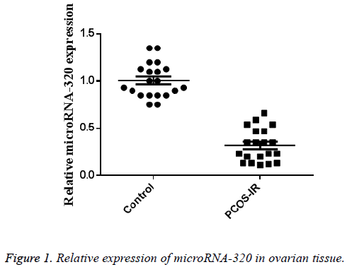
The ovarian tissues in 20 cases of patients with PCOS and IR (PCOS-IR group) and 20 cases of normal persons (control group) were collected. The total RNA of ovarian tissue was extracted. The RT-PCR results showed that, compared with the control group, the expression of microRNA-320 in PCO-IR group was significantly decreased (P<0.01), which was 0.35 × 0.24 times than that of the control group (n=20, P<0.01). The results are shown in Figure 1.
MicroRNA-320 targeted IRS-1 3’ UTR
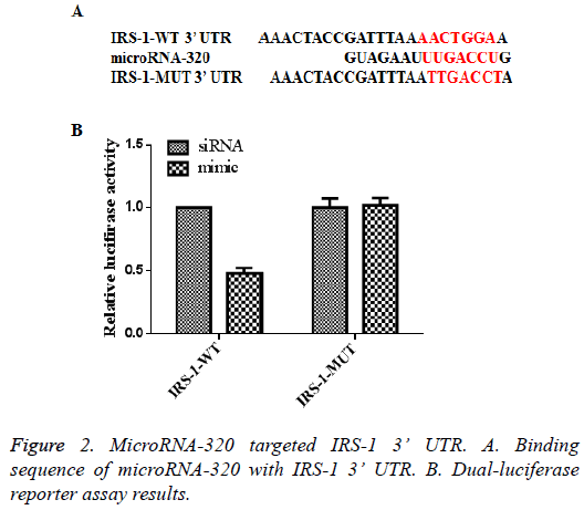
The bioinformatic software TargetScan and miRanda were used to predict the possible specific binding target sites with microRNA-320. Among them, the target gene IRS-1 was directly related to IR in patients with PCOS. The specific binding IRS-1 target sites with microRNA-320 are shown in Figure 2. The dual-luciferase reporter gene analysis showed that, compared with the control siRNA group, the activities of 293T cells transfected with mimic sequence and IRS-WT highexpression plasmid were significantly decreased. However, the activity of 293T cells luciferase containing mimic sequence and IRS-MUT did not significantly change. The results are shown in Figure 2. The results suggested that IRS-1 was a direct target of microRNA-320.
MicroRNA-320 inhibiting the expressions of IRS-1 and ERK/2
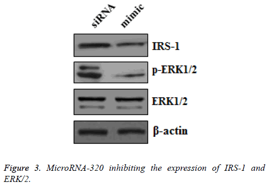
To further investigate the downstream signal pathways of microRNA-320 in inhibiting the targeted IRS-1, the 293T cell protein samples were collected after the transfected mimic sequence and siRNA were cultured for 48 h. The expressions of IRS-1, p-ERK1/2, and ERK1/2 in the two groups were detected by Western blot. The result showed that the expressions of IRS-1 and p-ERK1/2 were significantly downregulated in 293T cells transfected with mimic sequence, but the expression of ERK1/2 did not significantly change (Figure 3). This result suggested that microRNA-320 inhibited ERK1/2 pathway via IRS-1.
Expressions of IRS-1 and ERK1/2 in ovarian tissue of patients with PCOS combined with IR
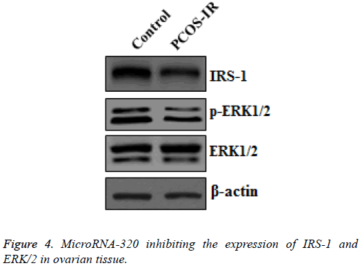
To investigate whether the same IRS-1-ERK1/2 signal pathway is present in ovarian tissue, the expressions of IRS-1, p- ERK1/2, and ERK1/2 in ovarian tissue of patients in PCOS–IR group and the control group were detected. Consistent with the results shown in Figure 3, compared with the low expression of microRNA-320 in the PCOS-IR group, the expressions of IRS-1 and p-ERK1/2 were highly expressed by Western blot, but the expression of ERK1/2 showed no significant change (Figure 4).
Discussion
Although microRNA research has been given considerable attention in recent years, few studies have focused on the expression and function of microRNA-320 in patients with PCOS. This study showed that the microRNA-320 expression in ovarian tissue showed significant difference in the PCOS-IR group, suggesting that microRNA-320 could regulate the IR in patients with PCOS. The complexity of microRNA network suggested that the expressions of microRNA showed difference between the patients with PCOS and patients without PCOS. Determination of the expression and function of some specific microRNAs in PCOS will provide a new target for the diagnosis and treatment of PCOS. The microRNA-320 might be regarded as a potential diagnostic and therapeutic target.
PCOS is often accompanied by IR in varying degrees and compensatory hyperinsulinemia, high androgens, and luteinizing hormone. Studies have shown that the incidence of IR in patients with PCOS is 50%-70% [6]. Further study on IR is of great significance in clarifying the pathogenesis of PCOS and in preventing complications. The serine/threonine phosphorylation of IRS protein is an important factor leading to IR. The IRS protein activity or its downstream signal could enhance the negative regulation of insulin signal transduction and its effect [7,8]. IRS-1 mainly activated the MAPK/ERK pathway. The RT-PCR result confirmed the low expression of microRNA-320 in patients with PCOS and predicted that IRS-1 might be a target gene of microRNA-320. The further dual-luciferase reporter gene method and Western blot confirmed the correlation between IRS-1 and microRNA-320.
IRS-1 could mediate the signal of insulin or insulin-like growth factor receptor and activate the signal pathway IR-related ERK1/2 [9,10]. This study showed that microRNA-320 could regulate the low expression of IRS-1 and then inhibit the phosphorylation of ERK1/2, which further revealed the important role of microRNA-320 in IR in PCOS.
Conclusion
In conclusion, our study suggested that microRNA-320 could regulate IRS-1 expression and ERK1/2 activity in ovarian tissue, which is associated with IR in PCOS. Our study contributes to the understanding of the mechanism of microRNA in PCOS combined with IR, thus providing theoretical basis for the diagnosis and treatment of PCOS.
References
- Schramm VG, Xavier B, Walter ME, Perlin VG, Motta LGJ, Cardoso LC, Dalmora SL. Analysis of insulin glargine in biopharmaceutical formulations by validated RP-LC and SE-LC Methods. Lat Am J Pharm 2017; 36: 332-339.
- Amr AEGE, Abdalla MM. Anticancer activities of some synthesized 2, 4, 6-trisubstituted pyridine candidates. Biomed Res India 2016; 27: 731-736.
- He A, Zhu L, Gupta N, Chang Y, Fang F. Overexpression of micro ribonucleic acid 29, highly up-regulated in diabetic rats, leads to insulin resistance in 3T3-L1 adipocytes. MolEndocrinol 2013; 21: 2785-2794.
- Zhao C, Yu T, Sun S, Wang W, Wu Y, Zhang Q. Glycyrrhizin antagonizes the hypoxia-induced chemoresistance of osteosarcoma cells. Lat Am J Pharm 2016; 35: 1199-1205.
- Ling HY, Hu B, Hu XB, Zhong J, Feng SD. MiRNA-21 Reverses high glucose and high insulin induced insulin resistance in 3T3-L1 adipocytes through targeting phosphatase and tensin homologue. ExpClinEndocrDiab 2012; 120: 553-559.
- Kaukab I, Nasir B, Abrar MA, Muneer S, Kanwal N, Shan SNH, Ahmad S, Murtaza G. Effect of pharmacist-led patient education on management of depression in drug resistance tuberculosis patients using cycloserine. A prospective study. Lat Am J Pharm 2015; 34: 1403-1410.
- Nteeba J, Bhattacharya P, Madden JA, Stromsdorfer K, Perfield JW. Effect of high fat diet-induced obesity on ovarian phosphatidylinositol-3 kinase signaling pathway members in mice. BiolReprod 2011; 85: 556-556.
- Liu GM, Xu K, Li J, Luo YG. 7, 8-dithydroxycoumarins protect human neuroblastoma cells from Aβ-mediated neurotoxic damage via inhibiting JNK and p38MAPK pathways. Biomed Res India 2016; 27: 591-595.
- Nteeba J, Ross JW, Keating AF. High fat diet induced obesity alters ovarian phosphatidylinositol-3 kinase signaling gene expression. ReprodToxicol 2013; 42: 68-77.
- Sailesh KS, Archana R, Sajeevan A, Mukkadan JK. Effect of controlled vestibular stimulation on depression, spatial and verbal memory scores in underweight female students-A pilot study. Biomed Res India 2016; 27: 611-615.


