ISSN: 0970-938X (Print) | 0976-1683 (Electronic)
Biomedical Research
An International Journal of Medical Sciences
Research Article - Biomedical Research (2017) Artificial Intelligent Techniques for Bio Medical Signal Processing: Edition-I
Biomechanical analysis of knee joint mechanism of the national women's epee fencing lunge movement
Fei Zhengwei1,2 and Zhao Chuanjie1,2*
1School of Physical Education and Sport Training, Shanghai University of Sport, Shanghai 200438, PR China
2Key Laboratory of Sport Skill and Tactic Diagnosis and Analysis General Administration of Sport of China, Shanghai 200438, PR China
- *Corresponding Author:
- Zhao Chuanjie
School of Physical Education and Sport Training
Shanghai University of Sport, PR China
Accepted date: January 14, 2017
Different movement patterns of fencing project are conducted through the right and left limbs, whose unique lunge attack defensive pace and great impact in the moment of the swing leg are considered as the main reasons to cause the swinging leg knee joint injury. How to reduce the injuries in knee lunge action has been a focus of research institutions at home and abroad. The application of sports biomechanics was analyzed in sports based on this. The biomechanical index of the National Women's epee reserve players lunge were tested considering fencing as an example, which was compared with dynamics index of no initial velocity falls naturally from 60 cm platform. Knee joint angle, ground reaction forces, knee joint torque and reaction force changes in the lunge movement of athletes were studied, and the causes of knee joint injury was analyzed. Practice has proved that the research of knee biomechanics mechanism of International Women's epee fencing lunge movement can improve the training effect and prevent sports injury effectively.
Keywords
Fencing, Lunge movemnent, Knee joint injury
Introduction
Fencing is a traditional project of the Olympic game. Competition is divided into men's foil, epee and sabre, women's foil, epee and sabre. Fencing is a skill that requires strength, speed, and power. Athletes control the movement of the body by step and control stalemate distance to opponent through the torso and the sword arm in the course of the competition in the field of athletes. They have a great time to attack when the opportunity to capture and conduct a sword attack opponents [1]. A lunge depth, speed and appropriateness are the key route of fencing to hit the opponent when observing favorable opportunity in the correct combat position [2]. The way of fencing is unique, and it is a project for a few of the left and right limbs to carry out different movement patterns. Its unique lunge attack defensive pace, swinging leg constantly braking and support are typical forms of emergency stop landing [3]. Lunge technique is one of the most important forms of fencing depth attack, which depends on the lunge action completed in almost all action scoring [4].
Sports biomechanics, as a subject of study on the structure and function of human body movement, plays an important role in sports training. Little et al. studied of fencing power quality characteristics and found that fencing strength quality is mainly wrist joints strength, shoulder and elbow joint strength, core strength, knee and ankle joint strength, arch elastic force according to joint with the analysis of isokinetic test on high level men's fencing athletes in China match kinescope and expert interviews. Among them, the key strength quality is the core strength, finger, wrist strength, leg strength [5]. Assari et al. investigated the ability of fencing athletes in the Olympic Games, and found that there was a significant difference between the lower limb muscle strength. At the same time, different types of fencing athletes also have obvious differences [6]. Haddad et al. studied the style of the same level, similar technology with a Fencing Club the sword in his right hand foilsman lunge distance, average speed of completion time, lunges and lunge top speed in the four case of lunges, which were in situ lunge, lunge step, jump lunge, step back lunge and after the jump lunge. The study found that the maximum average speed and the highest speed lunge were in a step after step jump lunges and lunge case, because getting an initial velocity through the ground reaction force in the process of retreat [7].
De et al. believed that the hind leg is significant to improve the speed of lunge and lunge velocity and angular velocity of the hock joint is closely related to the arrival time [8]. Kuntze et al. tested and analyzed before and after the lunge of leg of each joint angle speed, torque, power index to a high level of Fencing Athletes and found that the hind legs of knee and ankle joint power, torque and angular velocity were significantly greater than the forelegs of knee and ankle joints [9]. Sinclair and Bottoms discussed the main factors influencing the speed with lower extremity fencing lunge movement special features considering the biomechanical indexes of each joint in the future as the breakthrough point [10]. Possidente et al. studied choice of knee, ankle joint torque, angular velocity and joint angle index and analyzed the relevance of these indicators of lunge speed by using the grey correlation analysis method as well as finding the main influencing factors, which provided biomechanical basis for special strength training of Fencing Athletes [11]. This experiment aimed at the action of knee joint injury in fencing lunge was conducted, the lunge was conducted in detailed study and the dynamic indexes were analyzed by the method of biomechanics so that strive to reduce the athletes injury, improve the training effect. It is seen that quite a few work by Little et al. [5], Assari et al. [6], Haddad et al. [7], De et al. [8], Kuntze et al. [9] and Sinclair et al. [10] covers different aspects of training athletes. In this work, a biomechanical analysis of knee joint mechanism of the national women's epee fencing lunge movement is investigated.
Sports Biomechanics
Biomechanical analysis
Mechanics is a subject that studies the laws of mechanical motion of objects. That is the change of the position of object in time and space and the reason of its change. Biomechanics is applied to the mechanics of living organisms, which helps people to understand the normal function of the organ of life and its function, and predicts the change of its own change and puts forward the method of human intervention. Therefore, diagnosis, surgical operation and repair are all closely related to biomechanics. Sports biomechanics is a subject that studies the law of human body movement, and it is an important part of sports science. It reveals the biomechanical characteristics of the human body and the movement of the human body. It makes sports technology more reasonable and better. It improves the level of sports technology and performance and prevents damage and fitness guidance services (Figure 1).

Figure 1: Biomechanics of sports equipment improvement.
The role of sports biomechanics in sports
The research of sports biomechanics can improve the level of Sports Technology, which mainly displays in two aspects: Teachers and coaches apply mechanical knowledge to students and athletes to complete the exercise, improve the skill level (qualitative method), or biomechanics researchers have discovered new or more effective techniques to complete the project [12]. For example, students can’t complete before the roll and you can give two suggestions. One is the head of the chest and the body, and maintain in the rolling and another is to move forward over the top of the ground. The back style high jump technology was used in the Mexico Olympic Games in 1968. It can also improve the scientific of training. Technical analysis helps teachers and coaches identify places where students and athletes need to improve their training.
Sports biomechanics research can also improve sports equipment. As shown in the figure above, CAT-150 portable low oxygen tent can stimulate the body to speed up the formation of red blood cells, promote the flow of oxygen to the muscles and can let the body relax at athletes rest. VR goggles received feedback from the field. Players can judge the rowing is or not on the right direction with the help of this feedback in real time. Rowers can improve the formation of their ideal through real-time self-assessment. The design of the new MVA200 MiCasa limits the amount of sweat of volleyball to the palm so that the player has strengthened the control force to the ball. Embossed design seems not worth mentioning, which helps to reduce the resistance of air volleyball around so as to make the route more aerodynamic trajectory in volleyball. This will make the most difficult to pick the ball, but the sound is not so loud. The fourth generation of Speedo LZRRacer by the independent shark skin test that new swimsuit reduce the resistance by 4%, and can reduce 5% of oxygen consumption and performance improvement of 2% so that it become the world's fastest swimsuit and reached previously difficult to achieve buoyancy and sliding ability [13].
Biomechanical testing instrument
The main biomechanical test instrument has been widely used in sports, such as kinematic parameters-high speed camera, high speed video, three-dimensional force parameters, muscle EMG features, anthropometric parameters, model calculation, simulation etc. Three dimensional force measuring platform can reflect the reaction force of the ground to the human body. The strength of a player's ball is achieved through the interaction with the ground. Put pressure testing insoles into the shoes of the athletes can obtain sole pressure distribution in the process of athlete moving, which can provide effective parameters for the design of shoes for athletes [14]. EMG can track muscle activity intensity and time in the process of movement, which mainly involved in muscle. The athletes using EMG can know the completion of technical movement and muscle force, which provided an effective reference for training (Figure 2).

Figure 2: Sports biomechanical testing instruments.
Research Objects and Methods
Experiment object
A total of 6 women's epee athletes were selected in the National Fencing Team. One of them is international master, a national master and four national level athletes [15]. The researchers had no history of lower limb injury and participated in the experiment voluntarily. There is no muscle strain and joint strain in the recent months and the basic information of the subjects were as in Table 1.
| Athletes | age | height | weight | The sports life of athletes | Sports level | Dominant side |
|---|---|---|---|---|---|---|
| Li XX | 30 | 177 | 66 | 14 | International masters | right |
| Zhang XX | 28 | 179 | 64 | 12 | National athlete | left |
| Liu XX | 26 | 182 | 70 | 9 | Level of athletes | right |
| Zhan XX | 23 | 175 | 67 | 7 | Level of athletes | right |
| Wang XX | 28 | 174 | 66 | 10 | Level of athletes | left |
| Zhou XX | 29 | 180 | 71 | 11 | Level of athletes | left |
Table 1: The experimenter.
Experimental method
Experimental equipment: Infrared high speed imaging system, camera for 16 sets of MX13KISTLER three dimensional side force platform. It conducted connection and synchronization with VICON system through the digital analog converter and firmly placed in the measuring force platform pit. The age, height, weight, length of fencing, the level of athletes, the advantage side and the history of knee injury were recorded.
The VICON system was set up to adjust and calibrate the range of the shooting range before the experiment. The calibration ruler is shown in the Figure 3. Explain the experimental methods and attention to the subject and then fully warm up. Place in the receiver body infrared with reflective ball according to the requirements of the Visual3D modeling.

Figure 3: Epee lunges whereabouts and 60 cm high natural experiment.
The subjects were jogging for five minutes and then five minutes of stretching before the test. The subjects faced for the sword target and front and rear legs respectively were in the two block of force platform. The distance between sword and the subjects of the target was adjusted according to the receiver height so that the target distance athletes foot horizontal distance of 1.5 times the height of the toes. They asked the subjects with the fastest speed lunge target, each participant tries to stab 3 times and each time interval of 1 minutes of rest. 60 cm test platform was conducted after Bow thorn with three minutes and conducted no initial velocity under the condition of natural fall when feet to force platform, which was collected a total of 3 times (Figure 4). Attempt 3 times prior to the formal acquisition and the experiment member guided the subjects to realize the correct landing position.

Figure 4: Fencing epee competitions in the movement.
Data Processing and Analysis
Data processing
The pre-processed data in the VICON system was imported into the 3D Visual software. Kinematic data and GRF filter used low pass filtering. Construct the 14 parts of human body model in Visual 3D. Determine the body's center of gravity position according to the human body inertia parameters. Represent the speed to lunge in the horizontal direction of the body center of gravity speed. The three-dimensional kinematics and dynamics of the lower limb were calculated and derived in the 3D Visual software. Force, torque, knee flexion is negative (-) and stretching is positive (+). The starting point is defined as the lunge action front foot off the ground instantly (front GRF) and action endpoint is defined as the front knee extensor maximum angle.
Selection index
The kinetic parameters: Ground Reaction Force (GRF). Analyze ground reaction force on the Z axis and the X axis force and conduct the weight standardization. Knee joint moment: Decomposition in the calf coordinate system and the weight of standardization, the main data index is the Z axis, X axis and the direction of the force value. Joint reaction force: Decomposition in the calf coordinate system and the weight of standardization, the main indicators is the Z axis (compression force) and Y axis (shear stress) of the value and direction. The kinematics index is the knee joint flexion angle, which is composed of the big rotor, the lateral point of the knee and the step outside point to form the 3D angle.
Calculate the muscle torque of the joint of the human body or the binding force of the external and internal system of the human body according to the measurement of the various kinematic data for human motion [16,17].
Force analysis of lower limb joint
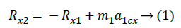
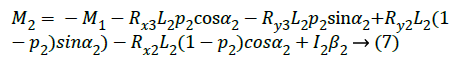
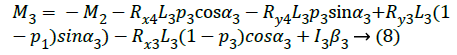
According to Newton's law and the moment of momentum of a single object, the equations of motion are listed. Taking the step of calculating the proximal joint from the far side of the limb, the reaction force and the joint muscle torque of all the joints are obtained. The detailed mathematical description is as follows:
The Rxi, Ryi (i=1,2,3,4), respectively, are the components of the binding force in the horizontal and vertical directions. Rx1 and Ry1 are the sole support reaction force, and the rest is the joint reaction force at the joint. M1, M2, M3 is respectively the net torque of the ankle, knee and hip joints [18]. For the inertia parameters of links, C1, C2, C3 represents the centroid of each link; Moment of inertia of the 3 aspects of their centroid axis is respectively I1, I2, I3; M1, M2, M3 is the quality of foot, leg and leg; P1, P2, P3 is the ratio of the distance from the centroid to the proximal end of each link and the length of the whole link. Components in the horizontal and vertical direction of acceleration of the center of mass of the other 3 links and link angular acceleration are a1cx, a2cx, a3cx, a1cy, a2cy, a3cy, ß1, ß2, ß3.The distance between the vertical support and the second toe joints is dc. The length of foot, leg and leg length is respectively L1, L2, L3, and the vertical angle is respectively α1, α2, α3 (Figure 5).


Then the statistics processing is carried on.

Figure 5: Lower limb joint force analysis diagram.
Research Results
Knee joint angle
The Table 2 shows, in a moment of swing leg landing, the ground reaction force, knee joint reaction force and knee joint torque almost reach the maximum at the same time. The size of the ground reaction force is closely related to the size of the joint reaction force and the joint moment. There are only 2 data with a deviation in the table. The knee angle of one subject with the maximum compressive force has a big difference with that when the maximum value of several dynamic indexes appear, and the difference is 8.4° and the interval time is 7% seconds. While the other deviation is with the maximum value of the knee joint moment, but the difference is not big with 10, and the interval time is only 1% seconds.
| The ground reaction force | Joint reaction force (compression force) | Joint reaction force (shear) | Joint torque |
|---|---|---|---|
| 19.6 | 19.6 | 19.6 | 19.6 |
| 23.2 | 23.2 | 23.2 | 23.2 |
| 29.6 | 29.6 | 29.6 | 30.6 |
| 12.5 | 20.9 | 12.5 | 12.5 |
| 24.0 | 24.0 | 24.0 | 24.0 |
| 26.0 | 26.0 | 26.0 | 26.0 |
Table 2: Ground, joint reaction force and moment of maximum peak corresponds to the knee joint Angle.
Ground reaction force
The vertical direction: In the Z axis direction of the ground reaction force (that is the vertical direction), three or more peaks are produced in the suffer landing of swing legs, and the first and third peaks are lager. The first peak is the largest with its force range between 2812.791 and 5674.49N, and the instantaneous value can reach 5.3 ± 1.3 times of body weight, as shown in Table 3.
| Swinging leg lunges stings the ground reaction force | |||
|---|---|---|---|
| The Z axis | The X axis | ||
| 5068.45 | 1683.23 | -423.23 | 1023.34 |
| 3787.32 | 1110.32 | -328.32 | 603.32 |
| 2813.32 | 966.23 | -492.23 | 332.12 |
| 5674.32 | 1023.32 | -309.21 | 829.32 |
| 4023.32 | 1153.23 | -667.43 | 276.32 |
| 4023.32 | 1173.32 | -668.32 | 276.32 |
| 3029.08 | 1149.32 | -767.32 | 535.23 |
Table 3: Lunges swinging leg when the peak ground reaction force.
Direction: It can be seen from Table 3, when the lunge with buffer landing, force generated by the ground reaction force is relatively small on the X axis than that of Z axis. However, all experimental data show that the direction of the ground reaction force in the early of landing stage is negative, with the basic duration percentage between 7% and 13% seconds, and the peak is between 325.33 N and 743.15 N. And in the following lunge landing process, the direction of the ground reaction force is changed into positive direction. Then compare the whereabouts of 60 cm in Table 4, its feature is contrast with that in the lunge-thrust.
| Swinging leg lunges stings the ground reaction force | |||
|---|---|---|---|
| The Z axis | The X axis | ||
| Peak 1 | Peak 2 or 3 | Peak 1 | Peak 2 |
| 1557.43 | 3558.23 | 983.32 | -97.32 |
| 3143.23 | 7344.23 | 1319.32 | -1313.10 |
| 1893.23 | 5374.32 | 685.21 | -806.23 |
| 1693.32 | 3996.22 | 713.23 | -672.32 |
| 3078.32 | 5382.21 | 1321.40 | -673.32 |
| 2759.32 | 7065.99 | 1196.75 | -894.32 |
Table 4: 60 cm tower of free fall the peak ground reaction force.
It can be seen from Table 5, in the lunge-thrust, knee joint will produce negative moment at the early landing of swing legs. Among them, the negative torque peaks of 4 subjects are between 298.16 Nm to 565.71 Nm. For the remaining 2 participants, there is the largest negative torque up to 1155.80Nm, and the smallest negative torque of the test staff has reaches 190.87 Nm. After impact, the torque direction of the experimental knee joint is turned into the positive moment. There are 3 people to maintain a positive moment until the end of the action, and the knee joint torques of the other 3 return to the negative torque, with the return into a positive moment until the end. Through the comparison of free fall landing from the 60 cm high platform with initial speed of zero, the maximum value of the knee joint torque range is from 332.55 Nm to 524.11 Nm. As can be seen from the Figure 6, the two kinds of test torque are different [19].

Figure 6: Lunges and 60 cm high test, ground reaction force.
| The peak torque (X) | |||
|---|---|---|---|
| In situ lunges thorn | 60 cm tower falling | ||
| Peak 1 | Peak 2 | Peak 1 | Peak 2 |
| -1155.80 | -165.08 | 162.06 | 411.10 |
| -190.87 | 112.05 | 171.75 | 400.10 |
| -298.16 | 21.49 | 148.37 | 332.55 |
| -365.64 | -113.4 | 148.08 | 332.11 |
| -565.71 | -83.83 | 119.73 | 524.11 |
| -385.78 | 35.92 | 284.07 | 450.25 |
Table 5: The peak torque (Nm).
Knee joint reaction force
It can be seen from Table 6, at the moment of the swing leg landing, tremendous counterforce of knee joint appears both in the direction of compression and shear stress. Huge compression force of the knee is in the range of 1077.37 N to 2675.73 N, the average equivalent to 2.1 ± 0.9 times of the weight, among which the maximum is up to 3.5 times of weight. The size of the shear force is close to the compressive force. The maximum shearing force value of the subjects is up to 2613.21 N, and has not much difference with maximum range of compression force. And it is found that the shear stress direction of the knee joint in the moment of impact is negative. Table 7 shows the knee high compression force generated by the whereabouts of the 60cm platform is greater than that of the lunge, the minimum compression force of 1768.67 N, and the maximum compressive force value reaches 6967.04 N. In the landing buffer process, the shear stress direction is the positive direction, which is different from the direction of shear stress generated when the swing legs are landing.
| Lunges swinging leg knee joint reaction force peak | |||
|---|---|---|---|
| The Z axis | The X axis | ||
| Peak 1 | Peak 2 | Peak 1 | Peak 2 |
| -2675.73 | -1310.07 | -2613.21 | 477.37 |
| -1194.19 | -1083.81 | -580.65 | 506.73 |
| -1225.93 | -1174.22 | -624.52 | 418.09 |
| -1077.37 | -1019.38 | -485.12 | 303.13 |
| -2149.96 | -1094.15 | -1321.42 | 314.57 |
| -1114.31 | -1143.25 | -862.40 | 339.31 |
Table 6: Lunges swinging leg knee joint reaction force peak (N).
| 60 cm peak tower falling knee joint reaction force | |||
|---|---|---|---|
| The Z axis | The X axis | ||
| Peak 1 | Peak 2 | Peak 1 | Peak 2 |
| -3062.25 | -1587.45 | 768.48 | 1369.79 |
| -6967.04 | -1698.60 | 1399.70 | 1396.00 |
| -4082.52 | -1383.37 | 787.60 | 1322.77 |
| -3404.67 | -1210.83 | 1301.03 | 1368.30 |
| -3704.78 | -1189.97 | 404.17 | 1996.28 |
| -1768.67 | -5019.73 | 1171.65 | 1303.12 |
Table 7: 60 cm peak tower falling knee joint reaction force (N).
Experimental biomechanical analysis
The maximum ground reaction force, knee joint moment and knee joint reaction force appear at the moment of heel landing in the fencing lunge swing leg, which shows that when the joint reaction force is maximum, the joint moment and ground reaction force are with the peak and there is a positive correlation between the three dynamic indexes. Though the comparison of ground reaction force and moment of 60 cm high free fall with zero initial velocity, great force and torque values endured by the knee joint in the swing leg’s landing for lunge-thrust are reflected in fact. And by comparison of the value of the reaction force in the vertical direction of the two landing actions, we find that both the minimum peaks of lunge-thrust and 60 cm landing have little difference. Fencing lunge-thrust in training and competition will be stride bigger and faster, and the ground reaction force is certainly far greater than the experimental data, thus increasing the knee joint’s bearing strength. It is easy to cause injury of the knee joint with the risk of fatigue or technology. The third peak of the lunge ground reaction force appears up to 1686.93 N, which also means that in the entire landing buffer process, the swing legs continue to withstand the impact of a huge ground reaction force, and the risk of knee injury may also increase.
In the comparison of the peak torque of knee joint in the two landing motions, we find that the peak moment of lunge of lunge-thrust and 60 cm high-profile landing are about in the same level, but there are 2 subjects over the 60 cm landing torque peak. It is worth mentioning that the lunge peak torque of one person exceeds about 3 times of his own 60 cm landing peak, and such a huge moment is a potential cause of knee injury. The huge lunge negative moment of knee joint is produced in the initial impact stage, indicating that before the swing leg landing, fencing athletes will make conscious active contraction of rear thigh muscles and active flexion and landing of shin. Longer time after soar of the swing leg not only is not conducive to continue to attack, but will become a good target for opponent to fight back. More stable landing with a shorter time as well as the quick transition of soar to lunge movement is more favorable to their own. However, at the moment of landing, the direction of a huge reaction is in anterior knee, indicating that the external torque direction generated by the ground reaction force is in the opposite direction with knee moment [20].
Lunge swinging leg also continues to bear a larger compression force in the moment of landing. Although the compression force is small compared to the whereabouts of the 60 cm platform, the average maximum compression force value has reached 2.1 ± 0.9 times as much weight. Therefore, for lunge-thrust, the compressive strength of the knee joint is still at a high level, and the whole process of action is showing a compression force until the end of the action. Joint reaction force continues to huge of larger compressive force direction, which shows that in the buffer stage of swing leg landing of the whole lunge-thrust, the knee joint bears sustained force. The force’s direction mainly acts in bone and cartilage, which is forced to squeeze between the bones, may cause cartilage inflammation of articular bones. Shear stress direction of swing leg lunge produced at the moment of landing is negative direction, indicating that the huge negative shear makes the backward displacement of tibia. In order to protect the tibia and prevent excessive displacement, posterior cruciate ligament will absorb a large number of shear forces to resist.
Conclusion
Fencing is a skill that requires strength, speed and power, and it is easy to cause sports injury in training and competition. Especially for the lunge action, the knee joint continues to bear the huge ground reaction force, which is easy to cause the injury of the knee joint. To solve this problem, the international women's lunge action is analyzed, and the mechanical index changes of natural fall of 60 cm with the zero initial velocity. The study finds that in the process of knee lunge, the knee will bear sustained force. The force’s direction mainly acts in bone and cartilage, which is forced to squeeze between the bones, may cause cartilage inflammation of articular bones, even possible stress fracture. The study also finds that in the buffer stage of swing leg in fencing lunge technique, mainly the rear-thigh muscle group endures external force. In future special training, it is suggested that the coaches pay attention to the muscle strength training of the rear-thigh muscle group, especially force training in the eccentric contraction of the rear-thigh muscle group. Experiments show that the research of knee biomechanics mechanism of epee fencing lunge movement helps to reduce the injury of athletes and provide theoretical basis for training, which is helpful to the development of fencing project in China. Similar kind of investigation can be carried out to assist the impact on knee joint in sports like marathon running, boxing, high jump and so on.
Acknowledgment
The work presented in this paper is supported by research base of Shanghai University of Sport; by the key technology research and application of the national fencing team preparing for the 2016 Rio Olympic Games (2013A032) and by the key players' application of skills and tactics preparing for the 2016 Rio Olympic Games (2016HT072).
References
- Moore KC, Chow FME, Chow JYH. Novel Lunge Biomechanics in Modern Sabre Fencing. Procedia Eng 2015; 112: 473-478.
- Greenhalgh A, Bottoms L, Sinclair J. Influence of surface on impact shock experienced during a fencing lunge. J Appl Biomech 2013; 29: 463-467.
- Gefen A, Shalom R, Elad D, Mandel Y. Biomechanical analysis of the keratoconic cornea. J Mech Behav Biomed Mater 2009; 2: 224-236.
- Swan KG Jr, Baldini T, McCarty EC. Arthroscopic suture material and knot type: an updated biomechanical analysis. Am J Sports Med 2009; 37: 1578-1585.
- Little KJ, Riches PE, Fazzi UG. Biomechanical analysis of locked and non-locked plate fixation of the clavicle. Injury-Int J Care Injured 2012; 43: 921-925.
- Assari S, Darvish K, Ilyas AM. Biomechanical analysis of second-generation headless compression screws. Injury-Int J Care Injured 2012; 43: 1159-1165.
- Haddad SL, Dedhia S, Ren Y, Rotstein J, Zhang LQ. Deltoid ligament reconstruction: a novel technique with biomechanical analysis. Foot Ankle Int 2010; 31: 639-651.
- de Jesus K, de Jesus K, Figueiredo P, Gonçalves P, Pereira S. Biomechanical analysis of backstroke swimming starts. Int J Sports Med 2011; 32: 546-551.
- Kuntze G, Mansfield N, Sellers W. A biomechanical analysis of common lunge tasks in badminton. J Sports Sci 2010; 28: 183-191.
- Sinclair J, Bottoms L. Gender differences in the kinetics and lower extremity kinematics of the fencing lunge. Int J Performance Anal Sport 2013; 13: 440-451.
- Possidente P, Phillips F, Matthis J. Anticipation of sabre fencing attacks. J Vision 2011; 11: 957-957.
- Zwierko T, Osinski W, Lubinski W. Speed of Visual Sensorimotor Processes and Conductivity of Visual Pathway in Volleyball Players. J Human Kinetics 2010; 23: 21-27.
- Feeley BT, Gallo RA, Sherman S, Williams RJ. Management of osteoarthritis of the knee in the active patient. J Am Acad Orthop Surg 2010; 18: 406-416.
- Keogh JW. Paralympic sport: an emerging area for research and consultancy in sports biomechanics. Sports Biomech 2011; 10: 234-253.
- Erdmann WS. Problems of Sport Biomechanics and Robotics. Int J Adv Robotic Syst 2013; 10: 323-330.
- Nicholls R, Fleisig G, Elliott B. Taylor & Francis Online: Baseball-Sports Biomechanics. Sports Biomechanics 2014.
- Howard RM, Conway R, Harrison AJ. A survey of sensor devices: use in sports biomechanics. Sports Biomech 2016; 15: 450-461.
- Chow JW, Knudson DV. Use of deterministic models in sports and exercise biomechanics research. Sports Biomech 2011; 10: 219-233.
- Geil MD. The Role of Footwear on Kinematics and Plantar Foot Pressure in Fencing. J Appl Biomechanics 2002; 18: 155-162.
- Roi GS, Bianchedi D. The science of fencing: implications for performance and injury prevention. Sports Med 2008; 38: 465-481.