ISSN: 0970-938X (Print) | 0976-1683 (Electronic)
Biomedical Research
An International Journal of Medical Sciences
Research Article - Biomedical Research (2017) Volume 28, Issue 1
Activity exerted by a naphthalene-oxirane derivative on the ischemia/reperfusion injury
1Laboratory of Pharmacochemistry, Faculty of Chemical and Biological Sciences, University Autonomous of Campeche. Av. Agustín Melgar, Col Buenavista C.P. 24039, Campeche Cam, México
2Escuela Nacional de Ciencias Biológicas del Instituto Politécnico Nacional. Prol. Carpio y Plan de Ayala s/n Col. Santo Tomas, México
3Facultad de Nutrición, Universidad Veracruzana. Médicos y Odontólogos s/n, 91010, Xalapa, Veracruz, México
- *Corresponding Author:
- Lauro Figueroa-Valverde
Faculty of Chemical and Biological Sciences
University Autonomous of Campeche
México
Accepted on April 01, 2016
There are data, which indicate that myocardial ischemia/reperfusion injury is a serious problem involved on cardiovascular diseases. Some naphtalen-derivatives have been used as cardioprotective drugs on myocardial ischemia-reperfusion injury; however, their activity and the molecular mechanism involved on myocardial ischemia-reperfusion injury are very confusing. Therefore, the aim of this study was to evaluate the activity of a naphtalen-oxirane derivative on myocardial ischemia/reperfusion injury using an isolated heart model. Additionally, molecular mechanism involved in the activity exerted by the naphtalen-oxirane derivative on perfusion pressure and coronary resistance was evaluated by measuring left ventricular pressure in absence or presence of following compounds; prazosin, metoprolol, indomethacin and nifedipine. The results showed that the naphtalen-oxirane derivative reduce infarct size compared with the control conditions. Other results showed that the naphtalen-oxirane derivative significantly increases the perfusion pressure and coronary resistance in isolated heart in comparison the control. Finally, other data indicate that the naphtalen-oxirane derivative increase left ventricular pressure in a dose-dependent manner; however, this phenomenon was significantly inhibited by nifedipine. In conclusion, all these data suggest that the naphtalen-oxirane derivative exert a cardioprotective effect through calcium channels activation and consequently induce changes in the left ventricular pressure levels. This phenomenon results in decrease of myocardial necrosis after ischemia and reperfusion.
Keywords
Naphtalen-oxirane derivative, Ischemia/reperfusion injury left ventricular pressure, Nifedipine.
Introduction
Several clinical and laboratory data indicate that myocardial infarction is a main cause of death worldwide [1,2]. In addition, there are studies that indicate that myocardial infarction can be induced by prolonged ischemia which consequently brings in cell viability, and ultimately cardiac function. Acute myocardial infarction can produce alterations in the topography of both the infarcted and non-infarcted regions of the ventricle [3]. There are some reports, which indicate the most effective method of limiting necrosis is restoration of blood flow; however, the effects of reperfusion itself may also be associated with tissue injury [4]. In the search of therapeutic alternatives to reduce the ischemiareperfusion injury, some drugs have been evaluated; for example, there is evidence that methylene blue decreases ischemia-reperfusion injury by inhibiting xanthine oxidase, which could bring as a consequence changes on superoxide concentration using an in vitro model of ischemia and reoxygenation [5]. Other data indicate that the compound 5- (N,N-dimethyl) amiloride induced protective effects on ischemia-reperfusion injury in rat hearts via inhibition of the Na+-H+ exchanger [6]. In addition, a study showed the protective effect of magnolol on ischemia-reperfusion injury attributed to its antioxidant and anti-inflammatory activity using a hind limb ischemic-reperfusion animal model [7].
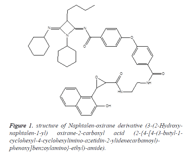
Other studies showed that glibenclamide reduces myocardial damage caused by ischemia/reperfusion in Guinea pig heart via activation of ATP-regulated K+ channels [8]. Also, other data indicates that Cyclosporin A can reduce the ischemia/ reperfusion-injury in isolated rat hearts through changes in cAMP levels [9]. In addition, some naphthalene derivatives have been developed to evaluate their biological activity in several animal models; for example, a study showed that the naphthalene derivative ((±)-(R,S)-5,6-dihydroxy-2- methylamino-1,2,3,4-tetrahydro-naphthalenehydro-chloride) can induces protective effects on ischemia-reperfusion injury in isolated heart rat via decrease of norepinephrine [10]. Other report, showed that the naphthalene derivative (N-(6- aminohexyl)-5-chloro-1-naphthalene sulfonamide), exerts cardioprotective effects on the isolated rat heart exposed to hypothermic and ischemic conditions by blocking the activity of calmodulin [11]. Other data indicates that compound 1,5- (dimethylamino)-N-(3,4-dimethyl-5-isoxazolyl)-1-naphthalene sulfonamide, significantly improved the recovery of cardiac function during reperfusion-ischemia injury through of reduced of creatine kinase (Figure 1) [12]. All these data show that naphtalene derivatives induce inotropic effects in the cardiovascular system; nevertheless, the cellular site and molecular mechanism involved in its inotropic activity are very confuse. Therefore, data information is needed to characterize the activity induced by the naphtalen derivatives at cardiovascular level. To provide this information, the present study was designed to investigate the effects of a naphtalen-oxirane derivative on perfusion pressure and vascular resistance in isolated rat hearts using the Langendorff technique. In addition, to evaluate the molecular mechanism involved in the inotropic activity induced by the naphtalen-oxirane derivative on left ventricular pressure the following compounds were used some pharmacological tools.
Material and Methods
All experimental procedures and protocols used in this investigation were reviewed and approved by the Animal care and use Committee of University Autonomous of Campeche (No. PI-420/12) and were in accordance with the guide for the care and use of laboratory animals [13]. Male Wistar rats; weighing 200-250 g were obtained from University Autonomous of Campeche.
Reagents
The compound 3-(2-Hydroxy-naphtalen-1-yl)oxirane-2- carboxyl acid (2-{4-[4-(3-butyl-1-cyclohexyl-4- cyclohexylimino-azetidin-2-ylidenecarbamoyl)- phenoxy]benzoylamino}-ethyl)-amide [14] was prepared with methods previously reported. In addition, another reagents used in this study were purchased from Sigma-Aldrich Co. Ltd. All drugs were dissolved in methanol and different dilutions were obtained using Krebs-Henseleit solution (≤ 0.01%, v/v).
Experimental design
Briefly, the male rat (200-250 g) was anesthetized by injecting them with pentobarbital at a dose rate of 50 mg/Kg body weight. Then the chest was opened, and a loose ligature passed through the ascending aorta. The heart was then rapidly removed and immersed in ice cold physiologic saline solution. The heart was trimmed of non-cardiac tissue and retrograde perfused via a non-circulating perfusion system at a constant flow rate. The perfusion medium was the Krebs-Henseleit solution (pH = 7.4, 37°C) composed of (mmol); 117.8 NaCl; 6 KCl; 1.75 CaCl2; 1.2 NaH2PO4; 1.2 MgSO4; 24.2 NaHCO3; 5 glucose and 5 sodium pyruvate. The solution was actively bubbled with a mixture of O2/CO2 (95:5/5 %). The coronary flow was adjusted with a variable speed peristaltic pump. An initial perfusion rate of 15 ml/min for 5 min was followed by a 15 min equilibration period at a perfusion rate of 10 ml/min. All experimental measurements were done after this equilibration period.
Perfusion pressure
Evaluation of measurements of perfusion pressure changes induced by drugs administration in this study were assessed using a pressure transducer connected to the chamber where the hearts were mounted and the results entered into a computerized data capture system (Biopac).
Evaluation of left ventricular pressure
Contractile function was assessed by measuring left ventricular developed pressure (LV/dP), using a saline-filled latex balloon (0.01 mm, diameter) inserted into the left ventricle via the left atrium [15]. The latex balloon was bound to cannula, which was linked to pressure transducer that was connected with the MP100 data acquisition system.
First stage
Ischemia/Reperfusion model: After of 15-minute equilibration time, the hearts were subjected to ischemia for 30 minutes by turning off the perfusion system [16]. After this period, the system was restarted and the hearts were reperfused by 30 minutes with Krebs-Henseleit solution. The hearts were randomly divided into 2 major treatment groups with n=9:
Group I. Hearts were subjected to ischemia/reperfusion but received vehicle only (Krebs- Henseleit solution). Group II. Hearts were subjected to ischemia/reperfusion and treated with naphtalen-oxirane derivative before ischemia period (for 10 minutes) and during the entire period of reperfusion. At the end of each experiment, the perfusion pump was stopped, and 0.5 ml of fluorescein solution (0.10%) was injected slowly through a sidearm port connected to the aortic cannula. The dye was passed through the heart for 10 sec to ensure its uniform tissue distribution. The presence of fluorescein was used to demarcate the tissue that was not subjected to regional ischemia, as opposed to the risk region. The heart was removed from the perfusion apparatus and cut into two transverse sections at right angles to the vertical axis. The right ventricle, apex, and atrial tissue were discarded. The areas of the normal left ventricle non risk region, area at risk, and infarct region were determined using methods previously reported [16]. Total area at risk was expressed as the percentage of the left ventricle.
Second stage
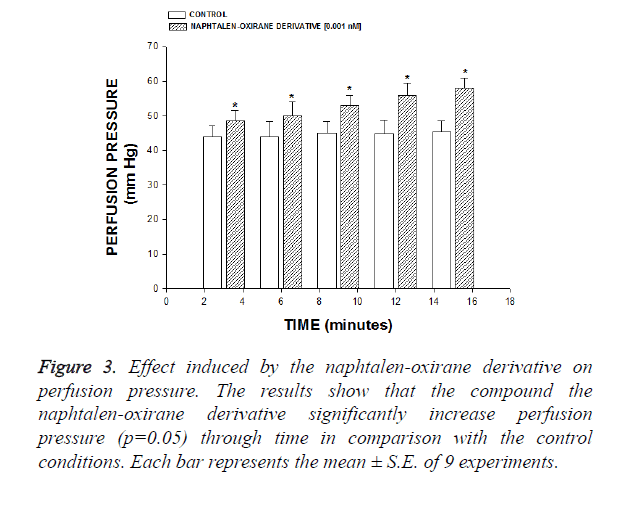
Effect induced by the naphtalen-oxirane derivative on perfusion pressure: Changes in perfusion pressure as a consequence of increases in time (3 to 18 min) in absence (control) or presence of the naphtalen-oxirane derivative at a concentration of 0.001 nM were determined. The effects were obtained in isolated hearts perfused at a constant-flow rate of 10 ml/min.
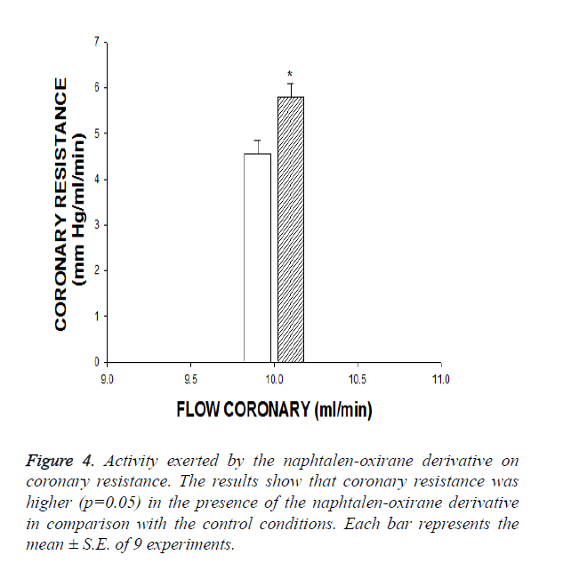
Evaluation of effects exerted by the naphtalen-oxirane derivative on coronary resistance: The coronary resistance in absence (control) or presence of estrone and the naphtalen-oxirane derivative at a concentration of 0.001 nM was evaluated. The effects were obtained in isolated hearts perfused at a constant flow rate of 10 ml/min. Since a constant flow was used changes in coronary pressure reflected the changes in coronary resistance.
Third stage
Effects induced by the naphtalen-oxirane derivative on left ventricular pressure through estrogen receptors: Intracoronary boluses (50 μl) of the naphtalen-oxirane derivative (0.001 to 100 nM) were administered and the corresponding effect on the left ventricular pressure was determined. The dose-response curve (control) was repeated in the presence of tamoxifen at a concentration of 1 nM (duration of preincubation with tamoxifen was by a 10 min equilibration period).
Effects induced by the naphtalen-oxirane derivative on left ventricular pressure through α1-adrenergic receptor: Intracoronary boluses (50 μl) of the naphtalen-oxirane derivative (0.001 to 100 nM) were administered and the corresponding effect on the left ventricular pressure was determined. The dose-response curve (control) was repeated in the presence of prazosin at a concentration of 1 nM (duration of preincubation with prazosin was by a 10 min equilibration period).
Effects induced by the naphtalen-oxirane derivative on left ventricular pressure through β1-adrenergic receptor: Intracoronary boluses (50 μl) of the naphtalen-oxirane derivative (0.001 to 100 nM) were administered and the corresponding effect on the left ventricular pressure was determined. The dose-response curve (control) was repeated in the presence of metoprolol at a concentration of 1 nM (duration of preincubation with metoprolol was by a 10 min equilibration period).
Effect exerted by the naphtalen-oxirane derivative on left ventricular pressure in the presence of indomethacin: The boluses (50 μl) of the naphtalen-oxirane derivative [0.001 to 100 nM] were administered and the corresponding effect on the left ventricular pressure was evaluated. The bolus injection administered was done in the point of cannulation. The dose response curve (control) was repeated in the presence of indomethacin at a concentration of 1 nM (duration of the pre-incubation with indomethacin was for a period of 10 min).
Effects of the naphtalen-oxirane derivative on left ventricular pressure through the calcium channel: Intracoronary boluses (50 μl) of naphtalen-oxirane derivative [0.001 to 100 nM] were administered and the corresponding effect on the left ventricular pressure was evaluated. The dose-response curve (control) was repeated in the presence of nifedipine at a concentration of 1 nM (duration of the pre-incubation with nifedipine was for a period of 10 min).
Effects induced by the naphtalen-oxirane derivative on the intracellular calcium levels: Changes in calcium levels as a consequence of increases in time (3-18 min) induces by the naphtalen-oxirane derivative (0.001 nM) were evaluated. The dose-response curve (control) was repeated in the presence of nifedipine at a concentration of 1 nM (duration of preincubation with nifedipine was by a 10 min equilibration period).
It is important to mention, that intracellular calcium was evaluated using the technique reported by Boe and Khan [17]. In this method, 5 mL of samples obtained of perfused (metabolic solution) were used to evaluate the intracellular calcium. It is important to mention that the samples were taken every 3 min (six times). After, 4 ml trifluoroacetic acid (10%) was added each to sample. This solution was mixed for 5 min at room temperature and after 1 mL of NaOH (25%) was added. To mixture 1 ml was trisodic phosphate added and the mixture was vortexed (1 min) and centrifuged to 4000 rpm (5 min). The supernatant was separated from the aqueous solution. After 5 mL of a buffer solution (pH=10) and 3 ml of eriochromeblack was added to the aqueous solution. The mixture was titled with Ethylene-Diamine-Tetra Acetic Acid (EDTA) (f=0.847).
Statistical analysis
The obtained values are expressed as average ± SE, using each heart (n=9) as its own control. The data obtained were put under Analysis of Variance (ANOVA) with the Bonferroni correction factor using the SPSS 12.0 program [18]. The differences were considered significant when p was equal or smaller than 0.05.
Results
First stage
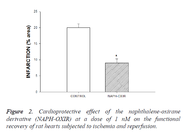
The results showed that the naphthalene derivative reduced infarct size (expressed as a percentage of the area at risk) compared with vehicle-treated hearts (Figures 2 and 3).
Figure 3. Effect induced by the naphtalen-oxirane derivative on perfusion pressure. The results show that the compound the naphtalen-oxirane derivative significantly increase perfusion pressure (p=0.05) through time in comparison with the control conditions. Each bar represents the mean ± S.E. of 9 experiments.
Second stage
In this study, the activity induced by a naphtalen-oxirane derivative on perfusion pressure and coronary resistance in isolated rat heart was evaluated. The results obtained from changes in perfusion pressure (Figure 3) as a consequence of increases in the time (3-18 min) in absence (control) or in presence of the naphtalen-oxirane derivative [0.001 nM], showed that this compound significantly higher the perfusion pressure (p=0.05) in comparison with the control conditions.
Third stage
Other result showed that coronary resistance, calculated as the ratio of perfusion pressure at coronary flow assayed (10 ml/ min) was higher in the presence of the naphtalen-oxirane derivative (p=0.05) than in control conditions at a concentration of 0.001 nM (Figure 4).
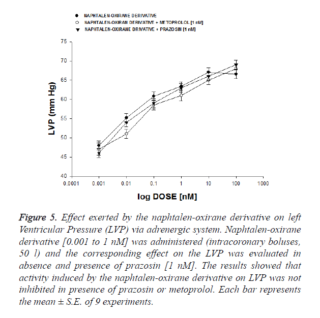
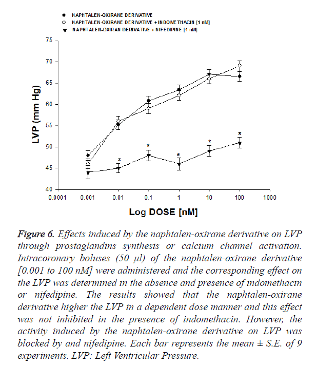
On the other hand, other experiments (Figure 5) showed that the naphtalen-oxirane derivative increase the perfusion pressure in a dose dependent manner (0.001 nM) and this effect was not inhibited in presence of prazosin (p=0.05) and metoprolol (p=0.06) at a concentration of 1 nM. Other results showed that the naphtalen-oxirane derivative increase the perfusion pressure in a dose dependent manner (0.001 to 1 nM) and this effect was not inhibited in presence of indomethacin (1 nM).
Figure 5. Effect exerted by the naphtalen-oxirane derivative on left Ventricular Pressure (LVP) via adrenergic system. Naphtalen-oxirane derivative [0.001 to 1 nM] was administered (intracoronary boluses, 50 l) and the corresponding effect on the LVP was evaluated in absence and presence of prazosin [1 nM]. The results showed that activity induced by the naphtalen-oxirane derivative on LVP was not inhibited in presence of prazosin or metoprolol. Each bar represents the mean ± S.E. of 9 experiments.
Other results showed that the activity exerted by the naphtalen-oxirane derivative (0.001 nM) on perfusion pressure (Figure 6) in presence of flutamide was not blocked (1 nM); however, nifedipine or finasteride significantly inhibited (p=0.05) the activity of the naphtalen-oxirane derivative at a concentration of 1 nM.
Figure 6. Effects induced by the naphtalen-oxirane derivative on LVP through prostaglandins synthesis or calcium channel activation. Intracoronary boluses (50 μl) of the naphtalen-oxirane derivative [0.001 to 100 nM] were administered and the corresponding effect on the LVP was determined in the absence and presence of indomethacin or nifedipine. The results showed that the naphtalen-oxirane derivative higher the LVP in a dependent dose manner and this effect was not inhibited in the presence of indomethacin. However, the activity induced by the naphtalen-oxirane derivative on LVP was blocked by and nifedipine. Each bar represents the mean ± S.E. of 9 experiments. LVP: Left Ventricular Pressure.
Finally, other results showed in the Table 1 indicate that activity induced by the naphtalen-oxirane derivative [0.001 nM] increased the intracellular calcium levels as a consequence of increases in the time (3-18 min); nevertheless, this effect is significantly reduced (p=0.005) in presence of nifedipine [1 nM].
| [Cai++] | ||
|---|---|---|
| Time (min) | Naphtale-oxirane | Naphtalen-oxirane + nifedipine |
| 3 | 1.44 × 10⁻³ | 2.00 × 10⁻⁴ |
| 6 | 1.44 × 10⁻³ | 2.00 × 10⁻⁴ |
| 9 | 1.44 × 10⁻³ | 2.00 × 10⁻⁴ |
| 12 | 2.00 × 10⁻³ | 2.00 × 10⁻⁴ |
| 15 | 2.00 × 10⁻³ | 2.00 × 10⁻⁴ |
Table 1. Activity induced by the Naphtalen-oxirane derivative [0.001 nM] on calcium intracellular [Cai++] in absence or presence of nifedipine [1 nM].
Discussion
Several drugs have been used to treat the infarction/reperfusion injury resulting from myocardial ischemia [19-21]; nevertheless, there is scarce information about of activity exerted by the naphtalene derivatives on this phenomenon. Therefore, in this study the activity of a naphtalen-oxirane derivative on the infarction/reperfusion injury was evaluated using several strategies.
First stage
In this step, the activity of the naphtalen-oxirane derivative was evaluated on injury by ischaemia/reperfusion using an isolated heart model. The results showed in the Figure 2 indicate that naphtalen-oxirane derivative reduced infarct size compared with vehicle-treated hearts (control). This phenomenon could be because the naphtalen-oxirane derivative exerts some influence on blood pressure, which could result reduction in the infarct size, and decrease the myocardial injury after ischemia-reperfusion.
Analyzing these data, in this study the activity induced by the naphtalen-oxirane derivative on blood vessel capacity and coronary resistance; translated as changes in perfusion pressure was evaluated in an isolated rat heart model. The results showed that the naphtalen-oxirane derivative significantly increase the perfusion pressure over time compared with the control. This phenomenon could be via activation of some structure biological (p.e. ionic channels or specific receptors) involved in the endothelium of coronary artery [22] which might result as changes on coronary resistance. To evaluate this hypothesis, the effect induced by the naphtalen-oxirane derivative on coronary resistance was evaluated. The results showed that coronary resistance in presence of this compound was higher in comparison the control conditions.
Analyzing these results and other data which indicate that some substances can exert effects on coronary resistance and left ventricular pressure [23]; in this study, the activity of the naphtalen-oxirane derivative on left ventricular pressure was evaluated. The results showed that compound the naphtalen-oxirane derivative increase the left ventricular pressure in a manner dose dependent. These data indicated that this compound might activate some bioactive substance (ionic channels or specific receptors) which could result in changes in left ventricular pressure.
In the search of molecular mechanism involved in the effect exerted by the naphtalen-oxirane derivative on the left ventricular pressure on left ventricular pressure; in this study, also were analyzed some reports, which indicates that some compounds can induce changes on left ventricular pressure via activation adrenergic receptors [24,25]. Therefore, in this study the activity exerted by the naphtalen-oxirane derivative on left ventricular pressure in the absence or presence of prazosin, metoprolol was evaluated. The results showed that the naphtalen-oxirane derivative increases the left ventricular pressure in a manner dose dependent and this effect was not inhibited by prazosin or metoprolol. These results indicate that the molecular mechanism involved in the effects of this compound on left ventricular pressure was not via adrenergic activity.
Analyzing these data and another reports which indicate that some drugs may induce its effect on left ventricular pressure via prostaglandins synthesis [26]; in this study the activity exerted by the naphthalene derivative on left ventricular pressure in the absence or presence of indomethacin was analysed to evaluate the possibility that the activities exerted by the naphthalene derivative involve stimulation and secretion of prostaglandins. The results showed that the naphthalene derivative increased the left ventricular pressure and this effect was not blocked in presence of indomethacin. These data indicate that activity exerted by the naphthalene-oxirane derivative on left ventricular pressure was not via prostanoids synthesis and secretion.
Evaluating all this data and reports, which indicate that some drugs exert their activity on left ventricular pressure through activation of calcium channel [27], in this study the effect exerted by the naphthalene-oxirane derivative on left ventricular pressure was evaluated using nifedipine as pharmacological tool to characterize whether this mechanism was involved in their pharmacological activity. The results showed that the naphthalene-oxirane derivative increases the left ventricular pressure in a dose dependent manner; however this effect was inhibited in presence of nifedipine. These data indicate that the activity exerted by the naphthalene-oxirane derivative was through activation of L-type calcium channels. To evaluate this hypothesis and analyzing other reports which indicate that activity induced by some compounds on left ventricular pressure involve an increase of intracellular calcium concentration [28]; In this study, the effect exerted by the naphthalene-oxirane derivative on intracellular calcium levels were evaluated in absence or presence of nifedipine. The results showed that, the effect exerted by the naphthalene-oxirane derivative induces increase in the intracellular calcium levels as a consequence of increases in the time; nevertheless, this effect is significantly reduced by nifedipine. Analyzing these data, the effect exerted by naphtalen-oxirane derivative on the calcium concentration was evaluated. The results showed that the naphtalen-oxirane derivative increased the calcium levels in comparison with control conditions.
Conclusion
The naphthalene-oxirane derivative is a particularly interesting drug, because the activity induced for this compound on injury by ischemia/reperfusion involves a molecular mechanism different in comparison with other drugs. This phenomenon may constitute a novel therapy for ischemia/reperfusion injury.
References
- Yusuf S, Hawken S, Ounpuu S. Obesity and the risk of myocardial infarction in 27 000 participants from 52 countries: a case-control study.Lancet 2005; 366: 640-649.
- Thygesen K, Alpert J. Universal definition of myocardial infarction. J Am CollCardiol 2007; 50: 2173-2195.
- Pfeffer M. Left ventricular remodeling after acute myocardial infarction. Ann Rev Med 1995; 46: 455-466.
- Przyklener K, Whittaker P. Deterious Effects of Oxygen Radicals in Ischemia/Reperfusion. Circulation 1989; 80: 1115-1127.
- Salaris C,Babbs C, Voorhees W. Methylene blue as an inhibitor of superoxide generation by xanthine oxidase: A potential new drug for the attenuation of ischemia/reperfusion injury. BiochemPharmacol 1991; 42: 499-506.
- Meng E, Pierce G. Protective effects of 5-(N,N-dimethyl)amiloride on ischemia-reperfusion injury in hearts. Am J Physiol-Heart 1999; 258: H1615-H1619.
- Chen H, Hung Y, Lee E. The protective efficacy of magnolol in hind limb ischemia-reperfusion injury. Phytomedicine 2009; 16: 976-981.
- McPherson C, Sontag D. ATP-regulated K+ channels protect the myocardium against ischemia/reperfusion damage. Circ Res 1991; 69: 571-581.
- Griffiths E, Halestrap A. ATP-regulated K+ channels protect the myocardium against ischemia/reperfusion damage. J MolCelCardiol 1992; 25: 1461-1469.
- Rossoni R. Manfredi B, Cavalca V.The Aminotetraline Derivative (±)-(R,S)-5,6-Dihydroxy-2-methylamino-1,2,3,4-tetrahydro-naphthalene Hydrochloride (CHF-1024) Displays Cardioprotection in Postischemic Ventricular Dysfunction of the Rat Heart.J PharmacolExpTher2003; 307: 633-639.
- Sugawara E, Nakayama Y, Senoo Y. Protective effects of calmodulin antagonists (trifluoperazine and W-7) on hypothermic ischemic rat hearts. Acta Med Okayama 1991; 45: 129-134.
- Tian-Lun Y, Mei-Fang C, Jun-Ling J. The endothelin receptor antagonist decreases ischemia/reperfusion-induced tumor necrosis factor production in isolated rat hearts. Int JCardiol 2005; 100: 495-498.
- Bayne, Revised Guide for the Care and Use of Laboratory Animals available. Am Physiol Soc. Physiologist 1996; 39: 208-211.
- Figueroa-Valverde L, Díaz-Cedillo F, Garcia-Cervera E. Design and synthesis of two new epoxides. Am J Chem 2015; 5.
- Figueroa-Valverde L, Díaz-Cedillo F, López-Ramos M. Inotropic Activity Induced by Carbamazepine-AlKyne Derivative in an Isolated Heart Model and Perfused to Constant Flow. Biomedica 2011; 31: 232-241.
- Booth E, Obeid N, Lucchesi B. Activation of Estrogen Receptor-α Protects the in Vivo Rabbit Heart from Ischemia-Reperfusion Injury. Am J Physiol 2005; 289: H2039-H2047.
- Boe J, Khan B. Colorimetric determination of blood calcium. J BiolChem 1929; 81: 1-8.
- Hocht C, Opezzo J, Gorzalczany S. UnaAproxi-maciónCinética y Dinámica de Metildopa en Ratas con CoartaciónAórticaMedianteMicrodiálisis. Rev ArgCardiol 1999; 67: 769-773.
- Node N, Kitakaze M, Kosaka H. Amelioration of Ischemia- and Reperfusion-Induced Myocardial Injury by 17β-Estradiol. Circulation 1997; 96: 1953-1963.
- Suparto I, Koudy W, Fox J. A comparison of two progestins on myocardial ischemia-reperfusion injury in ovariectomized monkeys receiving estrogen therapy. Coronary Artery Dis 2005; 16: 301-308.
- Jeanes H, Wanikiat P, Sharif I. A Medroxyprogesterone acetate inhibits the cardioprotective effect of estrogen in experimental ischemia-reperfusion injury. Menopause2006; 13: 80-86.
- Bouïs D, Hospers G, Meijer C. Endothelium in vitro: A review of human vascular endothelial cell lines for blood vessel-related research. Angiogenesis 2000; 4: 91-102.
- Figueroa-Valverde L, Diaz-Ku E, Diaz-Cedillo F. Effects of danazol and danazolhemisuccinate on perfusión pressure and vascular resistance. ActaBioquimClin Latin 2010; 44: 37-45.
- Esler S, Jennings G, Komesaroff P. Estrogen Supplementation Decreases Norepinephrine-Induced Vasoconstriction and Total Body Norepinephrine Spillover in Perimenopausal Women. Hypertension 1997; 30: 1538-1543.
- Peng N, Clark J, Wei C. Estrogen Depletion Increases Blood Pressure and Hypothalamic Norepinephrine in Middle-Aged Spontaneously Hypertensive Rats. Hypertension 2003; 41: 1164-1167.
- Seillan C,Ody C, Russo F. Differential aspects of sex steroids on prostaglandin secretion by male and female cultured piglet endothelial cells. Prostaglandins 1983; 26: 3-12.
- Figueroa-Valverde L, Diaz-Cedillo F, Lopez-Ramos M.Changes induced by estradiol-ethylenediamine derivative on perfusion pressure and coronary resistance in isolated rat heart:L-type calcium channel. Biomed Pap2011; 155: 27-32.
- Figueroa-Valverde L, Díaz-Cedillo F, García-Cervera E, Pool Gómez E, López-Ramos M, Rosas-Nexticapa M, Martinez-Camacho. Positive inotropic activity induced by a dehydroisoandrosterone derivative in isolated rat heart model. Arch Pharm Res 2013; 36: 1270-1278.
